 Back
BackChromosomal Variation: Structure, Number, and Cytogenetic Analysis
Study Guide - Smart Notes
 Tailored notes based on your materials, expanded with key definitions, examples, and context.
Microscopic Examination of Chromosomes
Cytogenetics and Chromosome Visualization
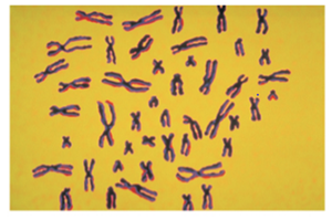
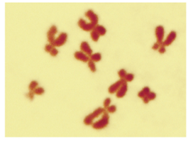
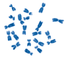
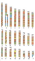
Cytogenetics is the field of genetics focused on the microscopic examination of chromosomes. Cytogeneticists analyze the chromosomal composition of cells to detect abnormalities in chromosome number or structure and to distinguish between species. Chromosomes are typically visualized during metaphase, when they are most condensed and visible under a microscope.
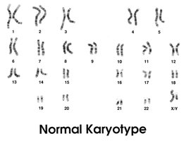
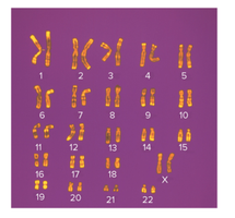
Karyotype: A micrograph in which all chromosomes of a single cell are arranged in a standard fashion, allowing for identification and comparison.
Applications: Detection of chromosomal abnormalities, species identification, and evolutionary studies.
Chromosome Classification Features
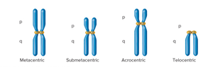
Chromosomes are identified and classified based on three main features:
Location of the centromere: Determines the chromosome's shape and classification (metacentric, submetacentric, acrocentric, or telocentric).
Size: Chromosomes vary in length, which aids in their identification.
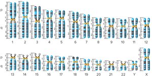
Banding patterns: Produced by staining techniques, such as G-banding, which reveal characteristic patterns of light and dark bands.
Karyotype Analysis
Karyotyping is a standard cytogenetic technique for arranging and analyzing the complete set of chromosomes in a cell. It is essential for diagnosing chromosomal abnormalities and for research in genetics and evolution.
Banding Patterns and Chromosome Identification
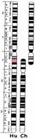
Banding patterns, such as those produced by Giemsa staining (G-banding), are crucial for distinguishing individual chromosomes, detecting structural changes, and studying evolutionary relationships. In humans, approximately 300 G bands are visible in metaphase and 800 in prometaphase.
Chromosome Structure and Evolution
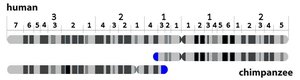
Chromosome 2 Fusion in Hominids
Comparative cytogenetics reveals that all members of the Hominidae family except humans have 24 pairs of chromosomes. Human chromosome 2 is the result of a fusion event between two ancestral chromosomes, which is supported by the presence of vestigial centromeres and telomeres.
Evidence: DNA sequence similarity between human chromosome 2 and two chimpanzee chromosomes.
Evolutionary significance: Demonstrates chromosomal rearrangement as a mechanism of speciation.
Changes in Chromosome Structure
Types of Structural Changes
Chromosome structure can be altered in two primary ways:
Change in total genetic material: Deletions (loss of segments) and duplications (repetition of segments).
Rearrangement without change in total material: Inversions (reversal of segment orientation) and translocations (segment attachment to a different chromosome).
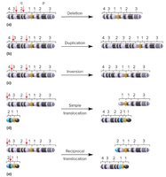
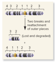
Deletions and Duplications
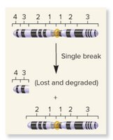
Deletions (deficiencies) occur when a chromosome breaks and a fragment is lost. The phenotypic consequences depend on the size and content of the deleted region. Large deletions are often detrimental, as seen in disorders like cri-du-chat syndrome (deletion on chromosome 5).
Terminal deletion: Loss of a chromosome end.
Interstitial deletion: Loss of an internal segment.
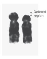
Cri-du-chat Syndrome
Cri-du-chat syndrome is caused by a deletion in the short arm of chromosome 5. It is characterized by severe mental retardation, physical abnormalities, and a distinctive cat-like cry in infants.
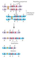
Duplications
Duplications are usually caused by abnormal recombination events, such as nonallelic homologous recombination. The phenotypic effects depend on the size of the duplicated region, but duplications are generally less harmful than deletions of similar size.
Gene Families and Evolution
Duplications can lead to the formation of gene families, groups of related genes derived from a common ancestor. Over time, duplicated genes may diverge in function, becoming paralogs within a species.
Example: The globin gene family, which encodes proteins for oxygen transport and storage, has evolved through multiple duplication events.
Copy Number Variation (CNV)
Copy number variation refers to segments of DNA that vary in copy number among individuals of a species. CNVs are common and can influence susceptibility to diseases such as schizophrenia, autism, and cancer.
Inversions and Translocations
Inversions
An inversion occurs when a chromosome segment is flipped in orientation. Most inversions do not affect phenotype, but they can cause problems if breakpoints disrupt vital genes (break point effect) or alter gene expression by changing gene position (position effect).
Pericentric inversion: Includes the centromere.
Paracentric inversion: Does not include the centromere.
Translocations
A translocation involves the transfer of a chromosome segment to a different chromosome. Reciprocal translocations exchange segments between two non-homologous chromosomes, usually without loss or gain of genetic material (balanced translocation). Simple (unbalanced) translocations involve the transfer in one direction and are often associated with phenotypic abnormalities.
Example: Familial Down syndrome is caused by a Robertsonian translocation between chromosomes 14 and 21.
Changes in Chromosome Number
Euploidy and Aneuploidy
Chromosome number can vary in two main ways:
Euploidy: Variation in the number of complete sets of chromosomes (e.g., diploid, triploid).
Aneuploidy: Variation in the number of particular chromosomes within a set (e.g., trisomy, monosomy).
Aneuploidy in Humans
Aneuploidy often leads to abnormal phenotypes due to gene dosage imbalance. The most common viable autosomal aneuploidies are trisomies 13, 18, and 21. Aneuploidies involving sex chromosomes are generally less severe due to mechanisms like X-inactivation.
Polyploidy and Its Consequences
Polyploidy (more than two sets of chromosomes) is common in plants and can result in larger, more robust individuals. In animals, polyploidy is usually lethal, but some tissues may be polyploid (endopolyploidy). Polyploid plants are important in agriculture, often producing seedless fruits and flowers.
Mechanisms Producing Chromosome Number Variation
Meiotic nondisjunction: Failure of chromosomes to segregate properly during meiosis, leading to gametes with abnormal chromosome numbers.
Mitotic nondisjunction: Occurs after fertilization, resulting in mosaicism (organism with genetically distinct cell populations).
Interspecies crosses: Hybridization between species can result in polyploid offspring.
Summary Table: Types of Chromosomal Structural Changes
Type | Description | Effect on Genetic Material |
|---|---|---|
Deletion | Loss of a chromosomal segment | Decreases |
Duplication | Repetition of a chromosomal segment | Increases |
Inversion | Segment reversed in orientation | Unchanged (rearranged) |
Translocation | Segment attached to a different chromosome | Unchanged (rearranged) |
