 Back
BackGenes, Proteins, Enzymes, and Human Disease: Molecular Genetics Study Guide
Study Guide - Smart Notes
 Tailored notes based on your materials, expanded with key definitions, examples, and context.
Genes and Human Disease
Historical Discovery of Genetic Diseases
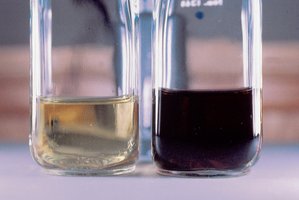
Mutations in genes can cause human diseases, as first described by Archibald Garrod in 1902 with the disease alkaptonuria. This disease is characterized by black urine and arthritis, resulting from the accumulation of homogentisic acid, which turns black upon exposure to air. Garrod termed this an 'inborn error of metabolism,' indicating a genetic rather than infectious origin.
Key Point: Alkaptonuria is inherited and not caused by germs.
Key Point: Garrod suggested it was a recessive Mendelian trait.
Example: The accumulation of homogentisic acid in urine is a direct result of a defective enzyme in the phenylalanine breakdown pathway.
What is a Gene?
Genetic and Biochemical Definitions
Genes have been defined both genetically and biochemically. Genetically, a gene controls some aspect of an organism’s phenotype and resides on chromosomes, segregating in defined ways. Biochemically, a gene is a segment of DNA containing the information to express a protein that performs a function in the cell or body.
Key Point: Genes are responsible for form, function, and behavior.
Key Point: Genes encode proteins, which are the functional molecules in cells.
Proteins: Structure and Function
Amino Acids and Protein Structure
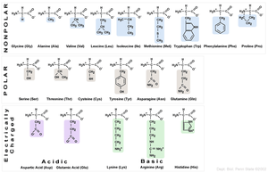
Proteins are polymers made of amino acids, each with a unique sequence. There are 20 amino acids commonly found in proteins, grouped by the chemical nature of their R side groups (nonpolar, polar, acidic, basic). The sequence and properties of these amino acids determine how a protein folds and functions.
Key Point: Proteins are linear chains of covalently joined amino acids.
Key Point: The bond between amino acids is called a peptide bond, formed by the ribosome.
Key Point: Polypeptide chains have an amino terminus (start) and a carboxyl terminus (end).
Example: The hemoglobin protein begins with a specific amino acid sequence.
Hydrophilic and Hydrophobic Amino Acids
The chemical nature of the R group determines whether an amino acid is hydrophilic (water-loving) or hydrophobic (water-fearing). Hydrophilic amino acids are generally found on the outside of proteins, exposed to water, while hydrophobic amino acids are found on the inside, sheltered from water.
Key Point: Amino acids are grouped by their R group properties.
Key Point: Protein folding is influenced by these properties.
Levels of Protein Structure
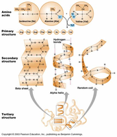
Proteins fold into precise three-dimensional structures based on their amino acid sequence. There are four levels of protein structure:
Primary structure: Linear sequence of amino acids joined by covalent bonds.
Secondary structure: Local structures formed by hydrogen bonds (alpha helix, beta sheet).
Tertiary structure: Final 3D shape formed by long-range interactions (hydrogen bonds, ionic bonds, hydrophobic/hydrophilic effects).
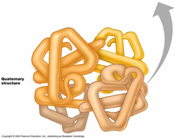
Quaternary structure: Multiple polypeptides working together (e.g., hemoglobin has 2 alpha and 2 beta subunits).
Genes, Proteins, and Enzymes
Enzymes as Proteins
Most genes contain instructions to make proteins, many of which are enzymes. Enzymes are proteins that catalyze specific chemical reactions, folding into shapes that allow them to perform these reactions at their active sites.
Key Point: Enzymes are proteins that perform chemical reactions.
Key Point: The active site is where the reaction occurs.
Example: Some enzymes break down molecules, others synthesize them.
Biochemical Pathways and Genetic Diseases
Alkaptonuria and Enzyme Defects
Alkaptonuria is caused by a defective enzyme in the pathway for breaking down phenylalanine, leading to the accumulation of homogentisic acid. Enzymes work together in pathways, and if one is missing, its substrate accumulates, causing disease.
Key Point: Enzyme defects can block biochemical pathways.
Key Point: Substrate accumulation is a hallmark of pathway defects.
Example: Alkaptonuria results from the inability to break down homogentisic acid.
Beadle and Tatum: One Gene-One Enzyme Hypothesis
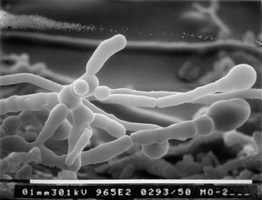
Beadle and Tatum used the bread mold Neurospora crassa to show that one gene controls one enzyme at a specific step in a biochemical pathway. They exposed spores to mutagens, isolated auxotrophic mutants, and determined which compounds the mutants could not synthesize.
Key Point: Auxotrophs cannot grow on minimal media and require supplements.
Key Point: Mutations in single genes affect specific steps in pathways.
Example: Mutants unable to synthesize methionine or arginine.
Human Genetic Diseases
Phenylketonuria (PKU)
PKU is caused by mutations in the gene encoding phenylalanine hydroxylase (PAH). Without PAH, phenylalanine accumulates and is broken down into harmful products, leading to severe mental retardation. Early detection and dietary management can prevent symptoms.
Key Point: PKU is a monogenic disease caused by enzyme deficiency.
Key Point: More than 400 recessive mutations in PAH are known.
Example: Newborns are screened for PKU and placed on a low-phenylalanine diet if affected.
Tay-Sachs Disease
Tay-Sachs disease is caused by a defect in the lysosomal enzyme HEXA, leading to substrate accumulation and neuronal degeneration. It is common among Jewish people of European ancestry and has no cure, but genetic testing has reduced its prevalence.
Key Point: HEXA deficiency causes substrate accumulation and neuronal damage.
Key Point: Tay-Sachs is a monogenic, heritable disease.
Sickle Cell Anemia
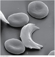
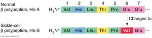
Sickle cell anemia is caused by mutations in the gene for beta hemoglobin. Hemoglobin is composed of two alpha and two beta chains. A single amino acid change (E6V: glutamic acid to valine) alters the protein’s structure, causing red blood cells to adopt a sickle shape, leading to fragility, blockage of capillaries, and reduced oxygen transport.
Key Point: Sickle cell anemia is a molecular disease caused by a single amino acid change.
Key Point: The mutation is common in populations exposed to malaria due to heterozygote advantage.
Example: Sickle cell carriers (heterozygotes) have increased resistance to malaria.
Protein Gel Electrophoresis and Molecular Disease
Electrophoresis in Sickle Cell Anemia
Linus Pauling showed that hemoglobin proteins from normal and affected individuals behave differently on a gel, allowing genotype determination by protein migration. The E6V mutation changes the charge and shape of hemoglobin, causing aggregation and sickling.
Key Point: Protein gel electrophoresis can distinguish wild type, carrier, and mutant hemoglobins.
Key Point: A single base pair change in DNA can cause a single amino acid change in a protein.
Sickle Cell Anemia and Malaria
Heterozygote Advantage
People heterozygous for the sickle cell mutation (HbA/HbS) have higher fitness in malarial environments. The presence of some mutant hemoglobin shortens RBC lifespan, disrupting the malaria parasite’s life cycle. This explains the high frequency of the sickle cell allele in populations exposed to malaria.
Key Point: Heterozygotes are less affected by malaria and do not have sickle cell disease.
Key Point: The sickle cell allele persists in malarial regions due to selective advantage.
Summary Table: Amino Acid Properties
The chemical nature of amino acid R groups determines their classification and role in protein structure.
Group | Amino Acids | Properties |
|---|---|---|
Nonpolar | Glycine, Alanine, Valine, Leucine, Isoleucine, Methionine, Tryptophan, Phenylalanine, Proline | Hydrophobic, found inside proteins |
Polar | Serine, Threonine, Cysteine, Tyrosine, Asparagine, Glutamine | Hydrophilic, found outside proteins |
Acidic | Aspartic acid, Glutamic acid | Negatively charged |
Basic | Lysine, Arginine, Histidine | Positively charged |
Key Equations and Concepts
Peptide Bond Formation:
Central Dogma:
One Gene-One Polypeptide Hypothesis: Each gene encodes a single polypeptide, which may be part of a larger protein complex.
Additional info: The notes cover topics from Ch. 4 (Gene Interaction), Ch. 10 (Eukaryotic Chromosome Abnormalities and Molecular Organization), Ch. 9 (The Molecular Biology of Translation), and Ch. 11 (Gene Mutation, DNA Repair, and Homologous Recombination), as well as applications in human disease genetics.
