 Back
BackMembrane Transport Proteins and Ion Channels: Structure, Function, and Genetic Relevance
Study Guide - Smart Notes
 Tailored notes based on your materials, expanded with key definitions, examples, and context.
Membrane Transport Proteins
Overview of Membrane Transport
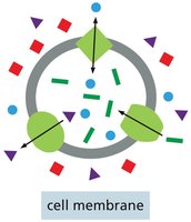
Membrane transport proteins are essential for the movement of molecules across the plasma membrane, which is otherwise impermeable to most substances. These proteins provide selective passageways for specific molecules and ions, maintaining cellular homeostasis and enabling critical biological processes.
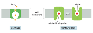
Transporters: Move small organic molecules or inorganic ions across the membrane by binding them specifically.
Channels: Form hydrophilic pores that allow substances to pass by diffusion, often discriminating based on size and charge.
Ion Channels: Permit passage of inorganic ions only.
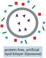
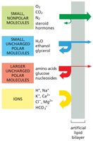
Permeability of Lipid Bilayers
Lipid bilayers are highly impermeable to ions and most uncharged polar molecules. The rate at which a molecule crosses a protein-free artificial lipid bilayer depends on its size and solubility.
Small, nonpolar molecules: Diffuse rapidly across membranes.
Uncharged polar molecules: Diffuse depending on size.
Charged molecules: Highly impermeable without transport proteins.
Ion Concentrations and Membrane Potential
Ion Distribution Across Membranes
Cells maintain distinct ion concentrations inside and outside the membrane, which is crucial for cellular function. The impermeability of cell membranes to inorganic ions allows for this regulation.
Extracellular: High Na+ balanced by Cl-.
Intracellular: High K+ balanced by negatively charged ions.
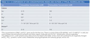
Table: Comparison of Ion Concentrations
Cation | Intracellular (mM) | Extracellular (mM) |
|---|---|---|
K+ | 5-15 | 145 |
Na+ | 5-15 | 150 |
Mg2+ | 0.5 | 1-2 |
Ca2+ | 10-4 | 1-2 |
H+ | 7 x 10⁻⁵ (pH 7.2) | 4 x 10⁻⁵ (pH 7.4) |
Cl- | 5-15 | 110 |
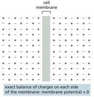
Membrane Potential
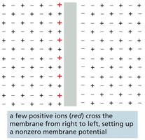
The voltage difference across the membrane, known as membrane potential, results from small excesses of positive or negative charge concentrated near the plasma membrane.
Resting membrane potential: -20 to -200 mV in animal cells.
Function: Powers transport of metabolites and is fundamental to electrical signaling.
Transporters and Channels
Types of Membrane Transport Proteins
Membrane transport proteins are highly selective, allowing passage of only specific molecules or ions.
Channels: Discriminate mainly by size and charge; open channels allow any suitable ion or molecule to pass.
Transporters: Bind solutes with high specificity, transferring only those that are specifically bound.
Passive and Active Transport
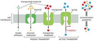
Solutes cross membranes by either passive or active transport.
Passive transport: Spontaneous movement down concentration gradient; includes all channels and some transporters.
Active transport: Movement against concentration gradient, requiring energy input; only performed by transporters (pumps).
Energy sources: ATP hydrolysis, ion gradients, sunlight.
Electrochemical Gradients and Osmosis
Electrochemical Gradient
The net force driving a charged molecule across a membrane is the electrochemical gradient, which combines the concentration gradient and membrane potential.
Determines direction: Each solute flows across a membrane by passive transport according to its electrochemical gradient.
Osmosis and Water Transport
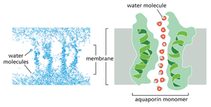
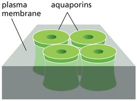
Water moves passively across cell membranes, but slowly. Specialized channels called aquaporins facilitate rapid water movement.
Osmolarity: Higher solute concentration inside cell pulls water in.
Osmosis: Movement of water down its concentration gradient.
Transporters: Specificity and Function
Transporter Specificity
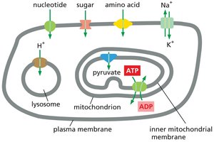
Transporters are responsible for moving most small, water-soluble organic and some inorganic molecules across cell membranes.
Highly selective: Often transfer only one type of molecule.
Cellular distribution: Each membrane contains a characteristic set of transporters.
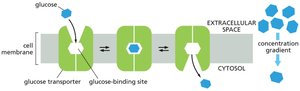
Passive Transporters
Passive transporters move solutes along their electrochemical gradient without determining direction.
Example: Glucose transporter moves glucose down its concentration gradient.
Active Transport and Pumps
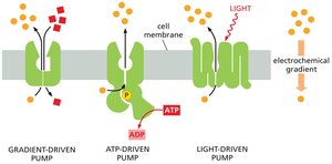
Types of Active Transport
Active transport is essential for maintaining correct intracellular solute concentrations.
Gradient-driven pumps: Couple uphill transport of one solute to downhill transport of another.
ATP-driven pumps: Hydrolyze ATP to drive transport.
Light- or redox-driven pumps: Use energy from sunlight or redox reactions.
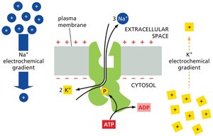
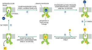
Na+-K+ Pump
The Na+-K+ pump uses ATP hydrolysis to pump Na+ out and K+ in, maintaining essential ion gradients.
Accounts for 30% of ATP consumption in animal cells.
Cycle: Each step depends on the previous, taking about 10 ms.
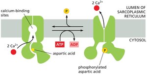
Ca2+ Pump
Ca2+ pumps keep cytosolic Ca2+ concentration low, which is critical for cellular signaling and muscle contraction.
ATPase: Similar to Na+ pump but does not require transporting a second ion.
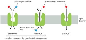
Gradient-Driven Pumps: Symport, Antiport, Uniport
Gradient-driven pumps use solute gradients to mediate active transport.
Symport: Moves both solutes in the same direction.
Antiport: Moves solutes in opposite directions.
Uniport: Moves only one solute.
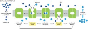
Glucose Transport and Electrochemical Gradients
Na+-Driven Glucose Transport
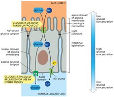
The electrochemical Na+ gradient drives the transport of glucose across the plasma membrane in animal cells.
Symport: Inward flow of Na+ powers glucose import even against its concentration gradient.
Cooperative binding: Both Na+ and glucose must bind for transport to occur.
Glucose Transporters in Gut Epithelium
Two types of glucose transporters enable gut epithelial cells to transfer glucose across the lining.
Passive glucose transporter: Releases glucose down its concentration gradient.
Active symport: Imports glucose from the gut.
Tight junctions: Keep transporters separated.
Ion Channels: Structure and Function
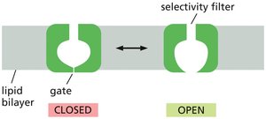
Ion Channel Selectivity and Gating
Ion channels are selective and gated, controlling which inorganic ions cross the membrane.
Selectivity filter: Determines ion specificity based on diameter, shape, and charge distribution.
Gating: Channels open briefly and close again, allowing controlled ion flow.
Transport rate: Channels transport ions >1000 times faster than transporters.
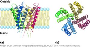
K+ Ion Channel Structure
The structure of K+ ion channels reveals the basis of ion specificity.
Channel width: Fits unhydrated K+ ions precisely.
Specificity: K+ passes 10,000 times more readily than Na+.
Membrane Potential and Ion Movement
Generation of Membrane Potential
Membrane potential is governed by the permeability of the membrane to specific ions.
Charge imbalance: Movement of ions sets up a voltage difference.
Small fraction: Only a small fraction of ions must move to establish membrane potential.
K+ Leak Channels and Resting Membrane Potential
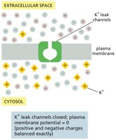
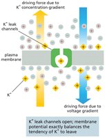
K+ concentration gradient and K+ leak channels play major roles in generating resting membrane potential in animal cells.
K+ leak channels: Open and close randomly, allowing K+ to flow out, leaving behind negative charges.
Voltage difference: Imbalance opposes further movement of K+.
Patch-Clamp Recording and Ion Channel Activity
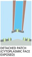
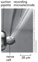
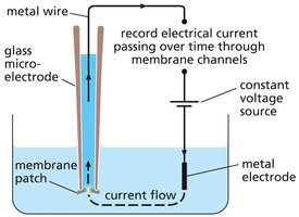
Patch-Clamp Technique
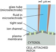
Patch-clamp recording is used to monitor ion channel activity by measuring electrical current through a single channel molecule.
Microelectrode: Isolates and makes electrical contact with a small area of membrane.
Applications: Determines ion specificity and gating behavior.
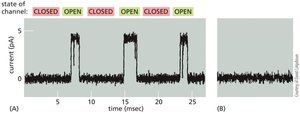
Ion Channel Activity: All-or-None Behavior
Ion channels randomly snap between open and closed states, exhibiting all-or-none activity.
Regulation: Conditions inside or outside the cell can bias channel behavior.
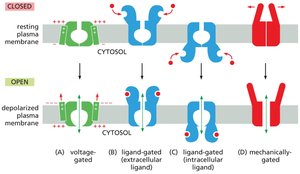
Types of Ion Channel Gating
Stimuli Influencing Ion Channel Opening
More than 200 types of ion channels exist, differing in ion specificity and gating mechanisms.
Voltage-gated: Opening influenced by membrane potential.
Ligand-gated: Controlled by binding of a ligand.
Mechanically-gated: Controlled by mechanical force.
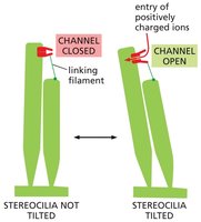
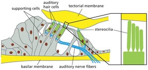
Mechanically-Gated Ion Channels in Hearing
Auditory hair cells in the ear depend on mechanically-gated channels.
Sound vibrations: Pull channel open, causing ions to flow into hair cells and generating electrical signals.
Voltage-Gated Ion Channels and Electrical Signaling
Voltage-gated ion channels respond to changes in membrane potential, enabling electrical signaling in cells.
Voltage sensors: Domains sensitive to membrane potential changes.
Control circuit: Fundamental to all electrical signaling in cells.
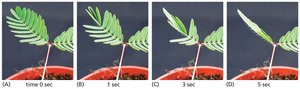
Plant Electrical Signaling: Leaf-Closing Response
Mechanically-gated and voltage-gated ion channels underlie the leaf-closing response in plants such as Mimosa pudica.
Touch stimulus: Opens mechanically-gated channels, triggering voltage-gated channels and electrical impulse.
Hinge cells: Rapidly lose water, causing leaflets to fold.
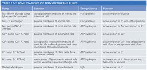
Summary Table: Examples of Transmembrane Pumps
Pump | Location | Energy Source | Function |
|---|---|---|---|
Na+-glucose symport | Kidney and intestinal cells | Na+ gradient | Active import of glucose |
Na+-H+ exchanger | Animal cells | Na+ gradient | Active export of H+, pH regulation |
Ca2+ pump (Ca2+ ATPase) | Eukaryotic cells | ATP hydrolysis | Active export of Ca2+ |
Na+ pump (Na+-K+ ATPase) | Animal cells | ATP hydrolysis | Active export of Na+ |
H+ pump (H+ ATPase) | Plant, fungi, bacteria | ATP hydrolysis | Active export of H+ |
Bacteriorhodopsin | Bacteria | Light | Active export of H+ |
Key Equations
Electrochemical Gradient
The electrochemical gradient () for an ion is given by:
R: Gas constant
T: Temperature
[C]_{in}, [C]_{out}: Ion concentrations inside and outside
z: Ion charge
F: Faraday constant
\Delta V: Membrane potential
Osmosis
Osmotic pressure () is given by:
c_{i}: Concentration of solute i
Conclusion
Membrane transport proteins and ion channels are fundamental to cellular physiology, genetic regulation, and signal transduction. Their specificity, gating mechanisms, and energy requirements are central to understanding cell biology and genetics. Additional info: These concepts are directly relevant to genetics, as membrane transport influences gene expression, cellular communication, and developmental processes.
