Textbook Question
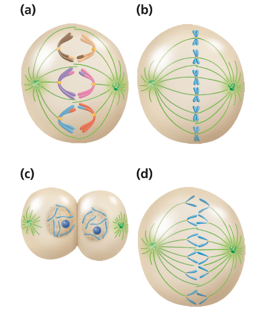
Our closest primate relative, the chimpanzee, has a diploid number of 2n = 48. For each of the following stages of M phase, identify the number of chromosomes present in each cell.
End of mitotic telophase
569
views

 Verified step by step guidance
 09:46 09:46
 07:10
07:10Our closest primate relative, the chimpanzee, has a diploid number of 2n = 48. For each of the following stages of M phase, identify the number of chromosomes present in each cell.
End of mitotic telophase
Our closest primate relative, the chimpanzee, has a diploid number of 2n = 48. For each of the following stages of M phase, identify the number of chromosomes present in each cell.
End of meiotic anaphase II
Our closest primate relative, the chimpanzee, has a diploid number of 2n = 48. For each of the following stages of M phase, identify the number of chromosomes present in each cell.
Meiotic metaphase I