Back
BackCHEM 1301 Exam 4 Study Guide: Biomolecules and Biological Chemistry
Study Guide - Smart Notes
 Tailored notes based on your materials, expanded with key definitions, examples, and context.
Chapter 20: Amino Acids and Proteins
Classification of Amino Acid Side Chains
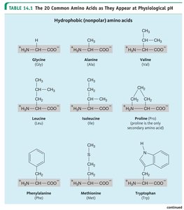
Amino acids are the building blocks of proteins and are classified based on the properties of their side chains (R groups): nonpolar (hydrophobic), polar (hydrophilic), acidic, or basic.
Nonpolar (hydrophobic) amino acids: Side chains are mostly hydrocarbons and do not interact favorably with water.
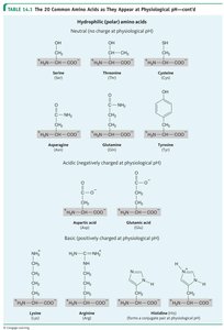
Polar (hydrophilic) amino acids: Side chains contain groups that can form hydrogen bonds with water.
Acidic amino acids: Side chains contain a carboxylic acid group and are negatively charged at physiological pH.
Basic amino acids: Side chains contain an amino group and are positively charged at physiological pH.
Example: Glycine is nonpolar; Glutamic acid is acidic; Lysine is basic.
Peptide Structure Analysis
Peptides are short chains of amino acids linked by peptide bonds. The N-terminus is the end with a free amino group, and the C-terminus is the end with a free carboxyl group.
Peptide bond: The amide linkage between the carboxyl group of one amino acid and the amino group of another.
Example: In the dipeptide Ala-Gly, Ala is at the N-terminus and Gly at the C-terminus.
Protein Structure Levels
Proteins have four levels of structure, each contributing to their function:
Primary structure: The linear sequence of amino acids in a polypeptide chain.
Secondary structure: Local folding patterns stabilized by hydrogen bonds (e.g., alpha helix, beta sheet).
Tertiary structure: The overall 3D shape of a single polypeptide chain, stabilized by interactions among side chains.
Quaternary structure: The arrangement of multiple polypeptide subunits in a protein.
Example: Hemoglobin has quaternary structure with four subunits.
Protein Denaturation
Denaturation is the loss of protein structure (and function) due to disruption of non-covalent interactions by heat, pH changes, or chemicals.
Causes: High temperature, extreme pH, detergents, heavy metals, organic solvents.
Effect: Loss of biological activity, but primary structure remains intact.
Enzyme Binding: Substrates and Inhibitors
Enzymes bind substrates at the active site. Inhibitors can bind at the active site (competitive) or at a different site (allosteric/noncompetitive).
Substrate: The molecule upon which an enzyme acts.
Inhibitor: A molecule that decreases enzyme activity by binding to the enzyme.
Chapter 21: Enzymes: Biological Catalysts
Role and Function of Enzymes
Enzymes are biological catalysts that speed up biochemical reactions by lowering the activation energy.
Six classes of enzymes: Oxidoreductases, Transferases, Hydrolases, Lyases, Isomerases, Ligases.
Example: Lactase catalyzes the hydrolysis of lactose.
Models of Enzyme Catalysis
Two main models describe enzyme-substrate interaction:
Lock and Key Model: The active site is a perfect fit for the substrate.
Induced Fit Model: The active site molds itself around the substrate upon binding.
Factors Affecting Enzyme Activity
Temperature: Enzyme activity increases with temperature up to an optimum, then decreases due to denaturation.
pH: Each enzyme has an optimal pH; deviations can reduce activity or denature the enzyme.
Substrate concentration: Activity increases with substrate concentration until the enzyme is saturated.
Enzyme Inhibition
Reversible inhibition: Inhibitor binds non-covalently and can dissociate.
Irreversible inhibition: Inhibitor binds covalently, permanently inactivating the enzyme.
Competitive inhibition: Inhibitor competes with substrate for the active site.
Noncompetitive inhibition: Inhibitor binds elsewhere, changing enzyme shape and reducing activity.
Enzyme Regulation
Allosteric control: Binding of a regulator at a site other than the active site alters enzyme activity.
Feedback control: The end product of a pathway inhibits an earlier enzyme in the pathway.
Cofactors and Vitamins
Cofactors: Non-protein molecules (metal ions or organic molecules) required for enzyme activity.
Vitamins: Many act as precursors to coenzymes.
Chapter 16: Carbohydrates
Classification of Sugars
Aldose: A monosaccharide with an aldehyde group (e.g., glucose).
Ketose: A monosaccharide with a ketone group (e.g., fructose).
Chirality and Anomeric Carbon
Chiral center: A carbon atom bonded to four different groups.
Anomeric carbon: The carbon derived from the carbonyl carbon (C=O) during ring formation; can be identified in Haworth projections.
Glycosidic Linkages
Glycosidic bond: The covalent bond joining two monosaccharides (e.g., β(1→4) linkage in lactose).
Reducing vs. Non-Reducing Sugars
Reducing sugar: Has a free anomeric carbon that can act as a reducing agent (e.g., maltose).
Non-reducing sugar: Both anomeric carbons are involved in glycosidic bonds (e.g., sucrose).
Glycogen Structure
Glycogen: A highly branched polysaccharide used for energy storage in animals.
Oxidation and Reduction of Sugars
Oxidation of aldose: Produces an aldonic acid.
Reduction of aldose/ketose: Produces sugar alcohols (alditols).
Chapter 18: Lipids
General Properties of Lipids
Lipids are hydrophobic biomolecules, including fatty acids, triglycerides, steroids, waxes, and soaps.
Fatty acid: Long hydrocarbon chain with a carboxylic acid group.
Triglyceride: Glycerol esterified with three fatty acids.
Steroid: Four fused hydrocarbon rings (e.g., cholesterol).
Wax: Ester of a long-chain fatty acid and a long-chain alcohol.
Soap: Salt of a fatty acid.
Triacylglycerols and Hydrolysis
Structure: Glycerol backbone with three fatty acids.
Hydrolysis: Produces glycerol and three fatty acids.
Saturation, Unsaturation, and Melting Point
Saturated fatty acids: No double bonds; higher melting point; usually solid at room temperature (fats).
Unsaturated fatty acids: One or more double bonds; lower melting point; usually liquid at room temperature (oils).
Cis vs. trans: Cis double bonds cause kinks, lowering melting point; trans double bonds behave more like saturated fats.
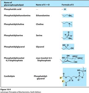
Glycerophospholipids and Head Groups
Glycerophospholipids are major components of cell membranes. The charge of a glycerophospholipid depends on the head group substituent.
Name of glycerophospholipid | Name of X—O | Formula of X |
|---|---|---|
Phosphatidic acid | — | H |
Phosphatidylethanolamine | Ethanolamine |
|
Phosphatidylcholine | Choline | — |
Phosphatidylserine | Serine | — |
Phosphatidylglycerol | Glycerol | — |
Phosphatidylinositol 4,5-bisphosphate | myo-Inositol 4,5-bisphosphate | — |
Cardiolipin | Phosphatidylglycerol | — |
Chapter 22: Nucleic Acids
Nucleotides, Nucleosides, and Nucleic Acids
Nucleoside: Nitrogenous base + sugar.
Nucleotide: Nitrogenous base + sugar + phosphate group.
Nucleic acid: Polymer of nucleotides (DNA or RNA).
Ribose vs. Deoxyribose
Ribose: Sugar in RNA; has a hydroxyl group at the 2' position.
Deoxyribose: Sugar in DNA; lacks the 2' hydroxyl group.
Pyrimidines and Purines
Pyrimidines: Cytosine, Thymine, Uracil (single ring).
Purines: Adenine, Guanine (double ring).
Watson-Crick Base Pairing
A-T: Two hydrogen bonds.
G-C: Three hydrogen bonds.
Phosphodiester Linkages and DNA Structure
Phosphodiester bond: Links nucleotides in a single strand.
Hydrogen bonds: Link complementary DNA strands.
DNA charge: Overall negative due to phosphate groups.
Complementary sequence: Each base pairs with its complement (A with T, G with C).
Chapter 23: RNA and Protein Synthesis
DNA vs. RNA Structure
DNA: Double-stranded, deoxyribose sugar, bases A, T, G, C.
RNA: Single-stranded, ribose sugar, bases A, U, G, C.
Stability of DNA Backbone
DNA backbone: More stable due to absence of 2' hydroxyl group, making it less susceptible to hydrolysis.
Ribosome Structure and Function
Ribosome: Composed of rRNA and proteins; site of protein synthesis (translation).
Transcription and Translation
Transcription: Synthesis of RNA from a DNA template.
Translation: Synthesis of protein from an mRNA template at the ribosome.
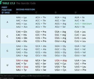
Genetic Code Table
The genetic code translates mRNA codons into amino acids. Each codon (three nucleotides) specifies an amino acid or a stop signal.
Mutations and Protein Structure
Mutation: A change in the DNA sequence that can alter protein structure and function.
Types: Silent, missense, nonsense, frameshift.
Appendices
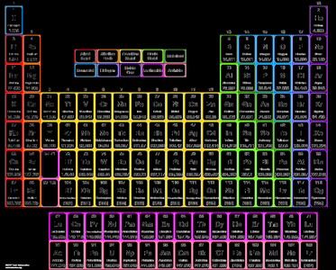
Periodic Table
The periodic table organizes elements by atomic number and properties. It is essential for understanding atomic structure, bonding, and chemical reactivity.