 Back
BackBIO5 Practical 2 Study Guide – Nervous System, Action Potentials, Eye Anatomy, and Endocrine System
Study Guide - Smart Notes
 Tailored notes based on your materials, expanded with key definitions, examples, and context.
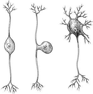
Q3. Be able to identify unipolar, bipolar, vs multipolar neurons (in a drawing or illustration)
Background
Topic: Structural Classification of Neurons
This question tests your ability to distinguish between the three main structural types of neurons based on their morphology.
Key Terms:
Unipolar neuron: One process extending from the cell body, typically found in sensory neurons.
Bipolar neuron: Two processes (one axon, one dendrite) extending from the cell body, found in special sensory organs.
Multipolar neuron: Multiple processes (one axon, many dendrites), most common in the CNS.
Step-by-Step Guidance
Examine the drawing and look for the number of processes extending from the cell body.
Identify the neuron with a single process (unipolar), two processes (bipolar), and multiple processes (multipolar).
Recall where each type is typically found: unipolar in sensory neurons, bipolar in special senses, multipolar in CNS and motor neurons.
Compare the structure in the drawing to these definitions to classify each neuron.
Try solving on your own before revealing the answer!
Final Answer:
From left to right: unipolar, bipolar, multipolar.
Unipolar has one process, bipolar has two, and multipolar has many dendrites and one axon.
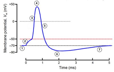
Q8. Describe the phases of action potential by referencing the diagram, mentioning the name of each phase, which specific ion channel is responsible, and which ion influx or efflux occurs.
Background
Topic: Action Potential Phases
This question tests your understanding of the sequence of events during an action potential, including the roles of specific ion channels and ion movements.
Key Terms and Formulas:
Resting membrane potential (RMP): The baseline electrical charge across the membrane.
Depolarization: Membrane potential becomes less negative due to Na+ influx.
Repolarization: Membrane potential returns toward RMP due to K+ efflux.
Hyperpolarization: Membrane potential becomes more negative than RMP.
Voltage-gated channels: Ion channels that open or close in response to changes in membrane potential.
Step-by-Step Guidance
Identify the starting phase (resting membrane potential) and note which channels are open or closed.
Describe the depolarization phase: opening of voltage-gated Na+ channels and Na+ influx.
Explain the peak of the action potential: inactivation of Na+ channels and opening of K+ channels.
Discuss repolarization: K+ efflux as voltage-gated K+ channels open.
Describe hyperpolarization: continued K+ efflux as channels remain open briefly.
Try solving on your own before revealing the answer!
Final Answer:
The phases are: 1) Resting membrane potential, 2) Depolarization, 3) Peak, 4) Repolarization, 5) Hyperpolarization, 6) Return to RMP.
Na+ influx causes depolarization, K+ efflux causes repolarization and hyperpolarization.
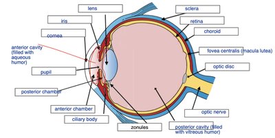
Q10. Identify the structures of the eye.
Background
Topic: Anatomy of the Eye
This question tests your knowledge of the anatomical structures of the eye and their locations.
Key Terms:
Cornea: Transparent front part of the eye.
Iris: Colored part controlling pupil size.
Lens: Focuses light onto the retina.
Retina: Contains photoreceptor cells.
Fovea centralis: Area of sharpest vision.
Optic nerve: Transmits visual information to the brain.
Step-by-Step Guidance
Study the labeled diagram and locate each structure: cornea, iris, lens, retina, fovea centralis, optic nerve, etc.
Note the function of each structure (e.g., cornea refracts light, lens focuses, retina detects light).
Identify the anterior and posterior chambers and their contents.
Observe the location of the optic disc and fovea centralis.
Try solving on your own before revealing the answer!
Final Answer:
The main structures are: cornea, iris, lens, retina, fovea centralis, optic disc, optic nerve, anterior and posterior chambers, ciliary body, sclera, choroid, zonules.
Each structure has a specific function in vision, as shown in the diagram.
