 Back
BackBody Organization and Medical Terminology: Foundations and Anatomical Reference
Study Guide - Smart Notes
 Tailored notes based on your materials, expanded with key definitions, examples, and context.
Body Organization
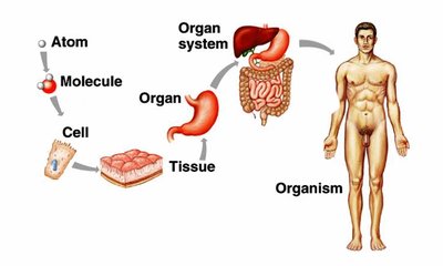
Levels of Body Organization
The human body is organized into hierarchical levels, each building upon the previous. Understanding these levels is essential for grasping medical terminology and the structure of the body.
Cells: The fundamental unit of life, capable of performing all basic life processes.
Tissues: Groups of similar cells working together to perform a specific function.
Organs: Structures composed of different types of tissues working together for a common purpose.
Systems: Groups of organs that perform complex functions necessary for survival.
Organism: The complete living individual, composed of all the above levels.
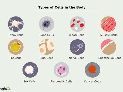
Cells
Cells are the smallest living units in the body and are responsible for all physiological processes. They:
Respond to stimuli
Engage in metabolic activity
Reproduce themselves
Form all tissues and organs
Specialized cells perform unique functions, such as muscle contraction or electrical impulse transmission.
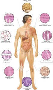
Tissues
Tissues are formed when similar cells group together to perform a specific activity. There are four primary types of tissue:
Muscular tissue: Produces movement by contracting.
Epithelial tissue: Forms protective barriers, lines organs, and is involved in absorption, secretion, and excretion.
Connective tissue: Supports and protects body structures; includes bone, cartilage, adipose, and tendons.
Nervous tissue: Conducts electrical impulses for communication throughout the body.
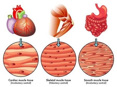
Muscular Tissue Types
There are three basic types of muscle tissue, each with distinct characteristics and locations:
Skeletal muscle: Attached to bones; responsible for voluntary movement.
Smooth muscle: Found in internal organs such as the intestines and uterus; involuntary control.
Cardiac muscle: Found only in the heart; involuntary control.
Anatomical Position and Body Planes
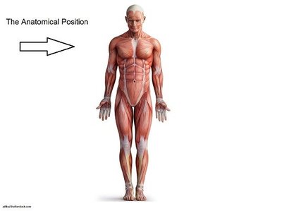
Anatomical Position
The anatomical position is the standard reference for describing locations and relationships of body parts. The body stands erect, arms at the sides, palms facing forward, eyes straight ahead, legs parallel, and feet pointing forward.
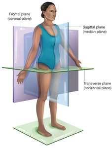
Body Planes
Body planes are imaginary lines used to divide the body for anatomical study and description. The three main planes are:
Sagittal (Median) Plane: Divides the body into left and right portions.
Frontal (Coronal) Plane: Divides the body into anterior (front) and posterior (back) portions.
Transverse (Horizontal) Plane: Divides the body into superior (upper) and inferior (lower) portions.
Body Regions and Cavities
Body Regions
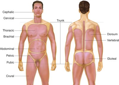
Specific terms are used to describe regions of the body for clarity in medical communication:
Brachial: Arm
Cephalic: Head
Cervical: Neck
Crural: Leg
Thoracic: Chest
Abdominal: Abdomen
Pelvic: Pelvis
Pubic: Genital region
Dorsum: Back
Vertebral: Spinal column
Gluteal: Buttocks
Body Cavities
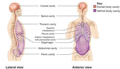
The body contains several cavities that house vital organs. These are divided into dorsal and ventral cavities:
Dorsal Cavities:
Cranial cavity: Contains the brain
Spinal cavity: Contains the spinal cord
Ventral Cavities:
Thoracic cavity: Contains the lungs and mediastinum (heart, aorta, esophagus, trachea, thymus gland)
Abdominopelvic cavity: Contains digestive, excretory, and reproductive organs; separated from thoracic cavity by the diaphragm
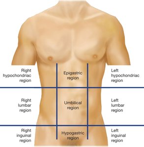
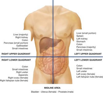
Anatomical and Clinical Divisions of the Abdomen
The abdomen is divided for anatomical and clinical reference:
Anatomical regions: Right/left hypochondriac, epigastric, right/left lumbar, umbilical, right/left inguinal, hypogastric
Clinical quadrants: Right upper (RUQ), left upper (LUQ), right lower (RLQ), left lower (LLQ)
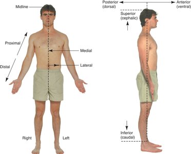
Descriptive Terms for Body Position
Directional Terms
Directional terms are used to describe the locations of structures relative to other structures or locations in the body:
Superior (Cephalic): Toward the head
Inferior (Caudal): Toward the feet
Anterior (Ventral): Toward the front or belly side
Posterior (Dorsal): Toward the back or spinal side
Medial: Toward the midline
Lateral: Toward the side
Proximal: Nearer to the point of attachment
Distal: Farther from the point of attachment
Apex: Tip or summit of an organ
Base: Bottom or lower part of an organ
Superficial: Closer to the surface
Deep: Further from the surface
Body Positions
Supine: Lying horizontally facing upward
Prone: Lying horizontally facing downward


Summary Table: Body Planes and Sections
Plane | Direction | Resulting Sections |
|---|---|---|
Sagittal (Median) | Vertical, front to back | Left and right portions |
Frontal (Coronal) | Vertical, side to side | Anterior and posterior portions |
Transverse (Horizontal) | Horizontal, parallel to ground | Superior and inferior portions |
