 Back
BackCardiovascular System: Medical Terminology and Anatomy Study Guide
Study Guide - Smart Notes
 Tailored notes based on your materials, expanded with key definitions, examples, and context.
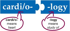
Cardiology: Introduction to Medical Terminology
Definition and Scope
Cardiology is the medical specialty focused on the study, diagnosis, and treatment of the cardiovascular system, which includes the heart and blood vessels. Understanding the terminology is essential for effective communication in healthcare settings.
Cardi/o-: Prefix meaning heart
-logy: Suffix meaning study of
Cardiology: The study of the heart and its functions
Cardiovascular System Overview
Structure and Function
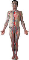
The cardiovascular system consists of the heart and an extensive network of blood vessels. It is responsible for circulating blood throughout the body, delivering oxygen and nutrients, and removing waste products.
Heart: Central organ that pumps blood
Blood vessels: Arteries, veins, and capillaries that transport blood
Continuous circulation: Ensures all body tissues receive oxygenated blood and nutrients
Circulation of the Blood
Pathways of Blood Flow
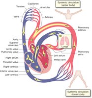
Blood circulates through two main pathways: pulmonary and systemic circulation. The heart acts as a double pump, sending deoxygenated blood to the lungs and oxygenated blood to the rest of the body.
Pulmonary circulation: Right side of the heart pumps deoxygenated blood to the lungs for oxygenation
Systemic circulation: Left side of the heart pumps oxygenated blood to the body
Key structures: Atria, ventricles, valves, arteries, veins, and capillaries
Anatomy of the Heart
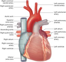
Location and External Features
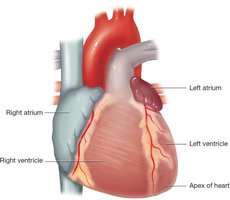
The heart is located in the thoracic cavity, behind the sternum and between the lungs. It is roughly the size of a fist and has an irregular external surface with mounds and grooves corresponding to internal chambers and vessels.
Chambers: Four chambers (right/left atria and right/left ventricles)
Grooves: Contain fat, blood vessels, and nerves
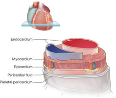
Heart Wall Layers
The heart wall is composed of three main layers, each with distinct functions:
Pericardium: Two-layered membrane forming the pericardial sac (parietal and epicardium layers)
Pericardial fluid: Lubricates and reduces friction between layers
Myocardium: Thick muscular layer responsible for contraction
Endocardium: Smooth inner lining of chambers and valves
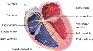
Internal Anatomy: Chambers and Septum
The heart contains four chambers separated by the septum:
Atria: Upper chambers that receive blood
Ventricles: Lower chambers that pump blood out
Septum: Central wall dividing right and left sides

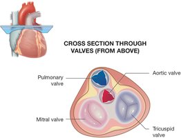
Heart Valves
Valves ensure unidirectional blood flow through the heart:
Tricuspid valve: Between right atrium and right ventricle
Pulmonary valve: Between right ventricle and pulmonary arteries
Mitral (bicuspid) valve: Between left atrium and left ventricle
Aortic valve: Between left ventricle and aorta
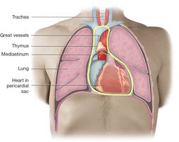
Anatomy: Thoracic Cavity and Mediastinum
Location of the Heart and Great Vessels
The heart is situated within the mediastinum, a central compartment in the thoracic cavity. This area also contains the great vessels, esophagus, trachea, and thymus.
Mediastinum: Central area between the lungs
Great vessels: Aorta, superior/inferior vena cava, pulmonary arteries/veins
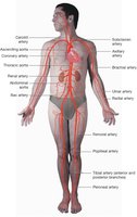
Anatomy: Blood Vessels
Types and Functions
Blood vessels are classified based on their structure and function:
Arteries: Carry blood away from the heart; thick muscular walls for high pressure
Capillaries: Smallest vessels; site of exchange between blood and tissues
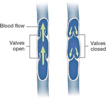
Veins: Carry blood toward the heart; thinner walls, often with valves
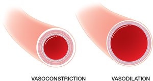
Arteries: Structure and Regulation
Arteries branch into smaller arterioles and are responsible for distributing oxygen-rich blood. They can constrict or dilate to regulate blood pressure.
Vasoconstriction: Narrowing of arteries, increases blood pressure
Vasodilation: Widening of arteries, decreases blood pressure
Major Arteries and Veins
The aorta is the largest artery, and the superior/inferior vena cava are the largest veins. Coronary arteries supply the heart muscle itself.
Veins: Structure and Function
Veins return deoxygenated blood to the heart. Many veins have valves to prevent backflow, especially in the limbs.
Venules: Small veins that collect blood from capillaries
Valves: Ensure one-way flow toward the heart
Circulation Pathways
Pulmonary and Systemic Circulation
Blood flows through two main circuits:
Pulmonary circuit: Right ventricle → pulmonary arteries → lungs → pulmonary veins → left atrium
Systemic circuit: Left ventricle → aorta → body tissues → veins → right atrium

Conduction System of the Heart
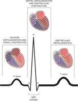
Electrical Pathways
The heart's rhythm is controlled by specialized cells that generate and conduct electrical impulses:
SA node (sinoatrial node): Pacemaker, initiates heartbeat
AV node (atrioventricular node): Delays impulse before passing to ventricles
Bundle of His, bundle branches, Purkinje fibers: Distribute impulse through ventricles
Physiology: Heartbeat and Regulation
Cardiac Cycle
The cardiac cycle consists of two main phases:
Systole: Contraction phase, blood is pumped out
Diastole: Relaxation phase, chambers fill with blood
Heart rate is regulated by the autonomic nervous system:
Parasympathetic: Releases acetylcholine, slows heart rate
Sympathetic: Releases norepinephrine and epinephrine, increases heart rate
Common Cardiovascular Diseases
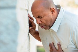
Angina Pectoris
Angina is chest pain caused by reduced blood flow to the heart muscle, often triggered by exertion or stress.
Peripheral Edema
Swelling in the lower legs and feet, often a sign of right-sided heart failure.

Myocardial Infarction (Heart Attack)
Occurs when blood flow to part of the heart is blocked, causing tissue death due to lack of oxygen.
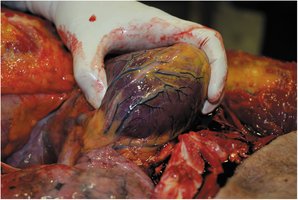
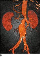
Aneurysm
A localized dilation or ballooning of a blood vessel wall, often in the aorta, which can rupture if untreated.
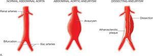
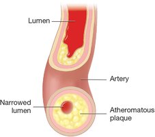
Atherosclerosis
Build-up of fatty plaques in arteries, narrowing the lumen and restricting blood flow.
Hypertension
Chronic high blood pressure, a major risk factor for heart disease and stroke.
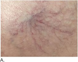
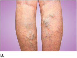
Varicose Veins
Enlarged, twisted veins, often in the legs, due to valve failure and blood pooling.
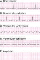
Arrhythmias
Abnormal heart rhythms, which can be detected on an electrocardiogram (ECG).
Bradycardia: Slow heart rate
Tachycardia: Fast heart rate
Fibrillation: Irregular, uncoordinated contractions
Asystole: Absence of heartbeat
Laboratory and Diagnostic Procedures
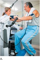
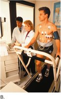
Cardiac Exercise Stress Test
Assesses heart function under physical stress using a treadmill or bicycle, monitoring ECG and blood pressure.
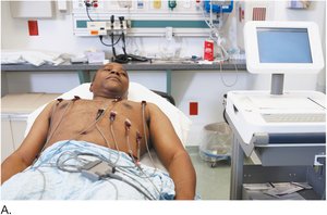
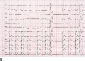
Electrocardiography (ECG/EKG)
Records the electrical activity of the heart to detect arrhythmias, ischemia, and other cardiac conditions.
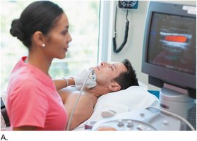
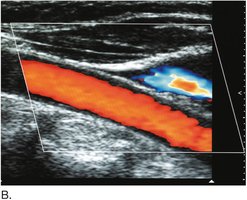
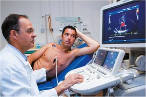
Ultrasonography and Echocardiography
Uses sound waves to visualize blood flow and heart structures. Color flow duplex ultrasonography shows both anatomy and blood flow direction/velocity.
Medical and Surgical Procedures
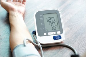
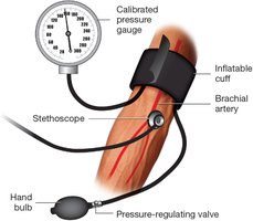
Measuring Blood Pressure
Blood pressure is measured using a sphygmomanometer and stethoscope. Systolic pressure reflects ventricular contraction; diastolic pressure reflects resting pressure.
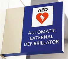
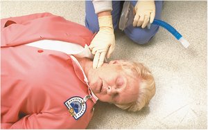
Automatic External Defibrillator (AED)
AEDs are portable devices used to treat sudden cardiac arrest by delivering an electric shock to restore normal rhythm.
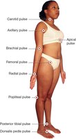
Pulse Points
Pulse points are locations where arterial pulses can be felt, used to assess heart rate and circulation.
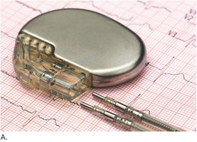
Pacemaker
A pacemaker is an implanted device that regulates heart rhythm in patients with arrhythmias.
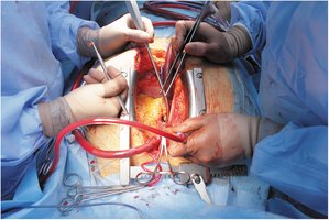
Open Heart Surgery
Involves opening the chest to access the heart for procedures such as valve replacement or coronary artery bypass.
Drugs and Pharmacology
Digitalis Drugs
Digitalis drugs, derived from the foxglove plant (Digitalis purpurea), are used to treat heart failure and arrhythmias by increasing the force of heart contractions.

Summary Table: Major Cardiovascular Terms
Term | Definition | Example/Application |
|---|---|---|
Cardiology | Study of the heart | Cardiologist diagnoses heart disease |
Myocardium | Heart muscle layer | Site of contraction |
Arrhythmia | Abnormal heart rhythm | Detected by ECG |
Angina | Chest pain from reduced blood flow | Occurs during exertion |
Aneurysm | Ballooning of vessel wall | Risk of rupture |
Hypertension | High blood pressure | Risk factor for stroke |
