 Back
BackCardiovascular System: Medical Terminology and Anatomy Study Guide
Study Guide - Smart Notes
 Tailored notes based on your materials, expanded with key definitions, examples, and context.
Cardiovascular System Overview
Functions and Major Organs
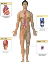
The cardiovascular system is responsible for distributing blood throughout the body, delivering essential substances to cells, and removing cellular waste. The main organs include the heart, arteries, capillaries, and veins.
Distribution: Blood is circulated to all body areas.
Delivery: Oxygen and nutrients are transported to cells.
Removal: Waste products are carried away for elimination.
Anatomy and Physiology of the Cardiovascular System
Blood Circulation
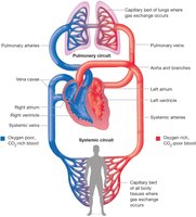
The cardiovascular system maintains blood distribution via two main circuits:
Systemic circulation: Carries blood from the heart to the body and back.
Pulmonary circulation: Carries blood from the heart to the lungs and back.
Heart Location and Structure
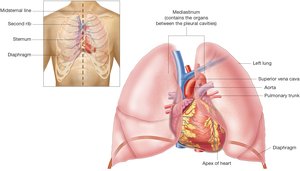
The heart is a muscular organ located in the mediastinum, about the size of a fist. It has four chambers and beats approximately 60–100 times per minute.
Location: Mediastinum of the thoracic cavity.
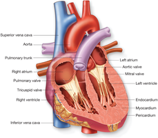
Chambers: Two atria (upper), two ventricles (lower).
Heart Layers
The heart wall consists of three layers:
Endocardium: Inner lining.
Myocardium: Muscular middle layer.
Pericardium: Protective outer layer.
Heart Chambers
The heart is divided into four chambers:
Atria: Receiving chambers (right and left).
Ventricles: Pumping chambers (right and left).
Septum: Divides right and left sides.
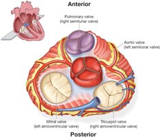
Heart Valves
Valves ensure unidirectional blood flow:
Tricuspid valve: Right atrium to right ventricle (three cusps).
Pulmonary valve: Right ventricle to pulmonary artery (semilunar).
Mitral valve: Left atrium to left ventricle (two cusps, bicuspid).
Aortic valve: Left ventricle to aorta (semilunar).
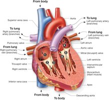
Blood Flow Through the Heart
Blood flows through the heart in a specific sequence:
Deoxygenated blood enters right atrium via vena cavae.
Blood moves to right ventricle through tricuspid valve.
Blood is pumped to lungs via pulmonary artery and valve.
Oxygenated blood returns to left atrium via pulmonary veins.
Blood moves to left ventricle through mitral valve.
Blood is pumped to body via aorta and aortic valve.
Diastole: Relaxation phase, chambers fill.
Systole: Contraction phase, chambers eject blood.
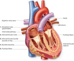
Conduction System of the Heart
The heart's contraction is regulated by its conduction system, controlled by the autonomic nervous system:
Sinoatrial (SA) node: Initiates electrical impulse, causing atria to contract.
Atrioventricular (AV) node: Receives impulse, transfers to AV bundle.
Bundle branches: Conduct impulse down the septum.
Purkinje fibers: Stimulate ventricles to contract.
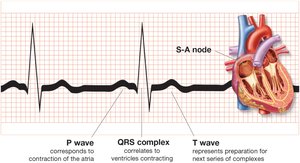
Electrocardiogram (ECG/EKG)
An electrocardiogram records the electrical activity of the heart:
P wave: Atrial contraction.
QRS complex: Ventricular contraction.
T wave: Preparation for next cycle.
Blood Vessels
Types of Blood Vessels
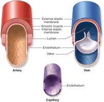
Blood vessels are classified into three types:
Arteries: Thick-walled, carry blood away from heart.
Capillaries: Tiny, thin-walled, site of exchange.
Veins: Thin-walled, carry blood toward heart, contain valves.
Lumen: Channel within vessel.
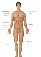
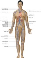
Major Arteries and Veins
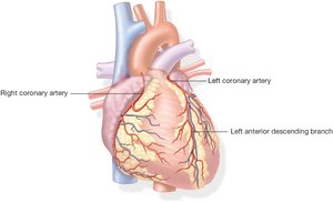
Key arteries include the aorta, pulmonary artery, and coronary arteries. Major veins include the superior and inferior vena cavae and pulmonary veins.
Capillaries
Capillaries connect arteries and veins, allowing for diffusion of oxygen, nutrients, carbon dioxide, and wastes.
Pulse and Blood Pressure
Blood pressure is the force exerted by blood against vessel walls, measured during:
Systole: Highest pressure (contraction).
Diastole: Lowest pressure (relaxation).
Factors affecting blood pressure include artery elasticity, vessel diameter, blood viscosity, and resistance.
Pulse is the surge of blood from heart contraction, typically measured at the wrist or throat.
Cardiovascular Pathology
Common Signs and Symptoms
Angiospasm: Involuntary muscle spasm in a vessel.
Ischemia: Reduced blood supply to tissues.
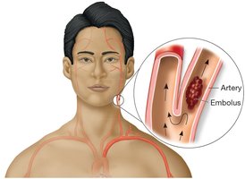
Embolus
An embolus is a blood clot or other substance that travels through the bloodstream and can lodge in a vessel, causing occlusion.
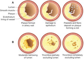
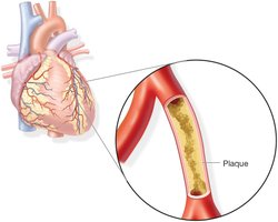
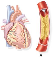
Atherosclerosis
Atherosclerosis is the buildup of plaque in arteries, leading to narrowing and potential blockage.
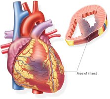
Myocardial Infarction
A myocardial infarction (heart attack) occurs when blood supply to part of the heart is blocked, causing tissue death (infarct).
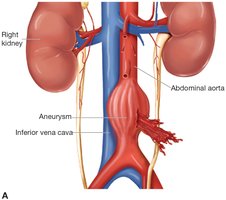
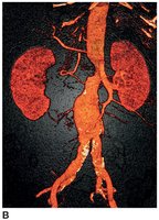
Aneurysm
An aneurysm is a localized dilation of a blood vessel, often in the abdominal aorta, which can rupture and cause severe bleeding.
Raynaud Phenomenon
Raynaud phenomenon is characterized by ischemic pallor of fingers, often triggered by cold.

Diagnostic and Therapeutic Procedures
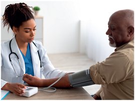
Blood Pressure Measurement
A sphygmomanometer is used to measure blood pressure.
Coronary Angiography
Angiography is the process of recording blood vessels, often used to assess coronary artery disease.
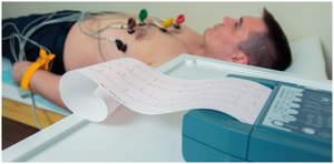
Electrocardiography and Monitoring
Electrocardiography: Records heart's electrical activity.
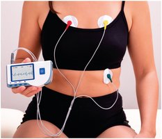
Holter monitor: Portable device for continuous ECG monitoring.
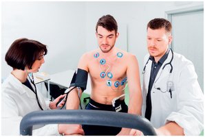
Stress test: Assesses cardiac function during exercise.
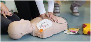
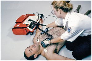
CPR and Defibrillation
CPR (Cardiopulmonary Resuscitation): Emergency procedure to restore circulation.
Defibrillation: Electrical shock to restore normal heart rhythm.
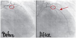
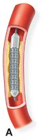
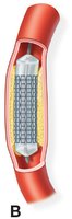
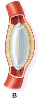
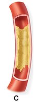
Stent Placement and Angioplasty
Stents and balloon angioplasty are used to treat blocked arteries:
Stent: Mesh tube placed to keep artery open.
Balloon angioplasty: Balloon compresses plaque against artery wall.
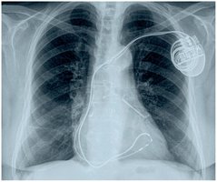
Pacemaker Implantation
A pacemaker is an electronic device implanted to regulate heart rhythm.
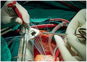
Prosthetic Valve Surgery
Prosthetic valves are surgically implanted to replace damaged heart valves.
Medical Terminology: Key Terms and Word Building
Combining Forms, Suffixes, and Prefixes
Combining forms: Root words used to build medical terms (e.g., cardi/o, angi/o).
Suffixes: Endings that modify meaning (e.g., -itis, -megaly).
Prefixes: Beginnings that modify meaning (e.g., endo-, extra-).
Examples
Cardiomegaly: cardi/o + -megaly = enlarged heart.
Endocarditis: endo- + cardi/o + -itis = inflammation of inner heart.
Thrombolytic: thromb/o + -lytic = destruction of a clot.
Extracorporeal: extra- + corpor/o + -eal = pertaining to outside the body.
Abbreviations
Common abbreviations in cardiovascular medicine include:
ECG/EKG: Electrocardiogram
BP: Blood pressure
CPR: Cardiopulmonary resuscitation
CAD: Coronary artery disease
Summary Table: Blood Vessel Types
Type | Structure | Function |
|---|---|---|
Artery | Thick-walled, elastic | Carry blood away from heart |
Capillary | Tiny, thin-walled | Exchange of gases and nutrients |
Vein | Thin-walled, valves | Carry blood toward heart |
Summary Table: Heart Valves
Valve | Location | Type | Function |
|---|---|---|---|
Tricuspid | Right atrium to right ventricle | Atrioventricular | Prevents backflow to right atrium |
Pulmonary | Right ventricle to pulmonary artery | Semilunar | Prevents backflow to right ventricle |
Mitral | Left atrium to left ventricle | Atrioventricular | Prevents backflow to left atrium |
Aortic | Left ventricle to aorta | Semilunar | Prevents backflow to left ventricle |
Key Equations
Blood Pressure Formula
Blood pressure is measured as:
Heart Rate Calculation
Pulse rate is generally equal to heart rate:
Conclusion
This guide summarizes the essential medical terminology, anatomy, physiology, pathology, and procedures related to the cardiovascular system, providing a comprehensive resource for exam preparation and clinical understanding.
