 Back
BackIntegumentary System and Dermatology: Medical Terminology Study Guide
Study Guide - Smart Notes
 Tailored notes based on your materials, expanded with key definitions, examples, and context.
Dermatology and the Integumentary System
Introduction to Dermatology
Dermatology is the medical specialty focused on the anatomy, physiology, diseases, and treatments of the integumentary system. This system includes the skin, nails, and subcutaneous tissue, and is essential for protection, sensation, and homeostasis.
Dermatology: dermat/o- means skin; -logy means study of.
Dermatologists diagnose and treat skin diseases using laboratory tests, medical procedures, and drugs.

Anatomy of the Integumentary System
Overview of Structures
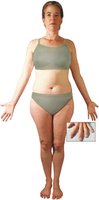
The integumentary system covers most of the body and consists of the skin, nails, and subcutaneous tissue. The skin is the largest organ in the body.
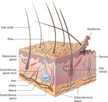
Skin Layers
The skin is composed of two main layers: the epidermis and the dermis. Beneath these lies the subcutaneous tissue.
Epidermis: Thin, outermost layer made of epithelial tissue. Contains living and dead cells, including melanocytes that produce melanin.
Dermis: Thick layer below the epidermis, made of connective tissue. Contains collagen, elastin, blood vessels, nerves, hair follicles, sebaceous glands, and sweat glands.
Subcutaneous tissue: Loose connective tissue containing adipose (fat) cells, providing insulation and protection.
Nails
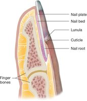
Nails are composed of both living and dead cells and consist of five parts: nail plate, nail root, lunula, cuticle, and nail bed.
Nail plate: Hard, flat part of the nail.
Nail root: Located under the skin, produces new nail cells.
Lunula: White, half-moon shape at the base.
Cuticle: Edge around the base of the nail.
Nail bed: Pink area under the nail plate.
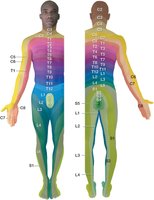
Dermatomes
A dermatome is an area of skin that sends sensory information to the spinal cord. Each dermatome is named according to the spinal nerve level it connects to (cervical, thoracic, lumbar, sacral).
Physiology of the Integumentary System
Protection
The skin acts as the body's first line of defense against injury and infection. Its acidic environment and keratinized cells discourage microorganism growth and make the skin waterproof.
Repair
When injured, the skin repairs itself through cell migration and division. Deep wounds form blood clots and scabs, while new cells fill in the damaged area.
Sensation
Sensory receptors in the dermis detect touch, pressure, vibration, pain, and temperature, sending signals to the nervous system for interpretation.
Vitamin D Synthesis
UV rays from the sun convert epidermal cholesterol into vitamin D, which is essential for calcium absorption. Sun exposure requirements vary by skin tone.
Thermoregulation
Thermoregulation is the process of controlling body temperature. Subcutaneous fat conserves heat, sweat glands cool the skin, and blood vessels adjust heat release.
Homeostasis
Homeostasis refers to the maintenance of internal balance. The integumentary system plays a key role in thermoregulation and signaling other medical conditions.
Diseases and Disorders of the Skin
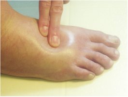
Edema
Edema is swelling caused by excess fluid in tissues. Pitting edema occurs when pressure leaves a deep indentation.
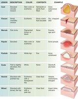
Types of Skin Lesions
Skin lesions are classified by their appearance, contents, and examples. Common types include cysts, fissures, macules, papules, pustules, scales, vesicles, and wheals.
Lesion | Description | Color | Contents | Example |
|---|---|---|---|---|
Cyst | Elevated circular mound | Skin color or erythema | Semisolid or partly fluid filled | Acne sebaceous cyst |
Fissure | Small, cracklike crevice | Erythema | None, some fluid exudate | Dry, chapped skin |
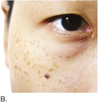
Macule | Flat circle | Pigmented brown or black | None | Freckle, age spot |
Papule | Elevated | Skin color or erythema | Solid | Acne pimple |
Pustule | Elevated | White top | Pus | Acne whitehead |
Scale | Flat to slightly elevated, thin flake | White | None | Dandruff, psoriasis |
Vesicle | Elevated with pointed top | Erythema with transparent top | Clear fluid | Herpes, chickenpox, shingles |
Wheal | Elevated with broad, flat top | Erythema with pale top | Clear fluid | Insect bites, urticaria |
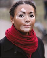
Skin Color Conditions
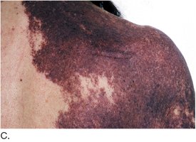
Skin color disorders include conditions such as albinism (lack of melanin), cyanosis (bluish skin due to lack of oxygen), necrosis (tissue death), and vitiligo (depigmentation).
Albinism: Normal number of melanocytes, but insufficient melanin production.
Cyanosis: Bluish discoloration, often seen in newborns or due to poor oxygenation.
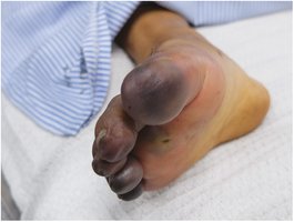
Necrosis: Tissue death from lack of blood supply, can lead to gangrene.
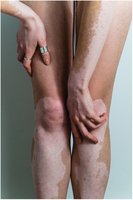
Vitiligo: Progressive autoimmune disorder causing depigmented patches.


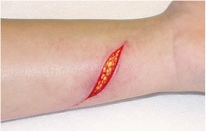
Skin Injuries
Common skin injuries include burns, keloids, lacerations, and pressure injuries.
Burns: Partial-thickness burns separate the epidermis from the dermis, causing blisters.
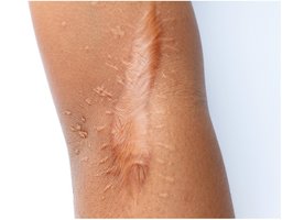
Keloid: Excessive scar tissue formation, larger than the original injury.
Laceration: Deep cut exposing subcutaneous tissue.
Pressure injury: Caused by prolonged pressure, leading to tissue damage.
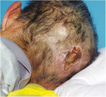
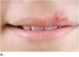
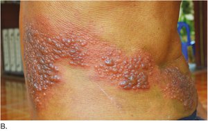
Skin Infections
Skin infections are caused by bacteria, viruses, or fungi. Examples include herpes simplex, shingles, and tinea capitis (ringworm).
Herpes simplex type 1: Vesicles on lips.
Shingles: Vesicles and crusts along a dermatome.
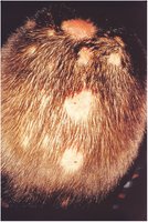
Tinea capitis: Fungal infection of the scalp, causing round lesions and hair loss.
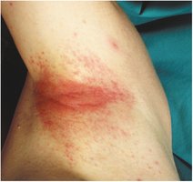
Allergic Skin Conditions
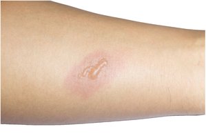
Allergic reactions can cause contact dermatitis, characterized by redness, irritation, and sometimes blistering.
Benign Neoplasms of the Skin
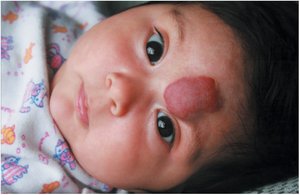
Benign skin growths include hemangiomas and nevi (moles). Nevi can be pigmented, flat, or resemble port-wine stains.
Hemangioma: Bright red lesion due to dilated blood vessels.
Nevus: Pigmented mole, can be raised, flat, or port-wine stain.
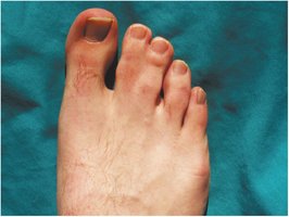
Congenital Skin Disorders
Syndactyly is a congenital condition where skin and soft tissues of fingers or toes are fused.
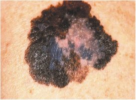
Malignant Neoplasms of the Skin
Malignant melanoma is a dangerous skin cancer characterized by asymmetry, irregular edges, varied color, and growth over time.
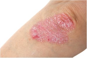
Autoimmune Disorders of the Skin
Psoriasis is a chronic autoimmune disorder causing red, scaly patches, commonly on elbows and knees.
Diseases of the Sebaceous Glands
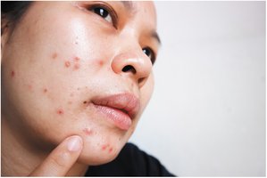
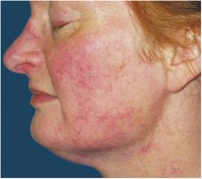
Acne vulgaris and rosacea are common disorders involving sebaceous glands.
Acne vulgaris: Papules and pustules triggered by increased sebaceous secretions during puberty.
Rosacea: Chronic erythema and dilated blood vessels, often affecting the face.
Laboratory, Diagnostic, Medical, and Surgical Procedures
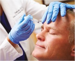
Botox Injection
Botox is a diluted neurotoxin used for cosmetic and medical purposes, such as reducing wrinkles.
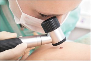
Skin Examination
Dermatologists use magnifying lenses to examine skin lesions, which may require biopsy for diagnosis.
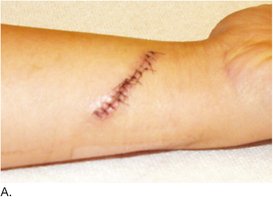
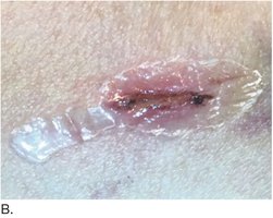
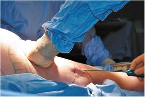
Wound Closure
Wounds can be closed with sutures or surgical glue. Sutures may be placed in layers, with deeper ones absorbed by the body.
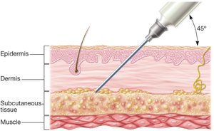
Hypodermic Injection
Hypodermic injections deliver drugs into the subcutaneous tissue, beneath the dermis and epidermis, at a 45-degree angle.
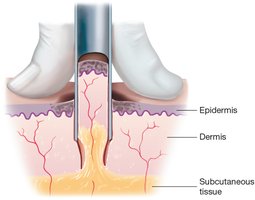
Punch Biopsy
A punch biopsy uses a circular cutter to remove a core of skin, including epidermis, dermis, and subcutaneous tissue, for diagnostic purposes.
Liposuction
Liposuction is a surgical procedure to remove fat from subcutaneous tissue, often for cosmetic reasons.
Summary Table: Common Skin Lesions
The following table summarizes the main types of skin lesions, their descriptions, colors, contents, and examples:
Lesion | Description | Color | Contents | Example |
|---|---|---|---|---|
Cyst | Elevated circular mound | Skin color or erythema | Semisolid or partly fluid filled | Acne sebaceous cyst |
Fissure | Small, cracklike crevice | Erythema | None, some fluid exudate | Dry, chapped skin |
Macule | Flat circle | Pigmented brown or black | None | Freckle, age spot |
Papule | Elevated | Skin color or erythema | Solid | Acne pimple |
Pustule | Elevated | White top | Pus | Acne whitehead |
Scale | Flat to slightly elevated, thin flake | White | None | Dandruff, psoriasis |
Vesicle | Elevated with pointed top | Erythema with transparent top | Clear fluid | Herpes, chickenpox, shingles |
Wheal | Elevated with broad, flat top | Erythema with pale top | Clear fluid | Insect bites, urticaria |
Key Medical Terminology
integument/o-: skin
cutane/o-: skin
melan/o-: black
-cyte: cell
pil/o-: hair
erect/o-: stand up
-ion: action or condition
Formulas and Equations
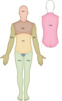
Rule of Nines for Burn Assessment
The Rule of Nines is used to estimate the percentage of body surface area affected by burns:
Head: 9%
Chest: 9%
Abdomen: 9%
Each arm: 9%
Genitals: 1%
Each leg: 18%
Back: 18%
Conclusion
The integumentary system is vital for protection, sensation, thermoregulation, and homeostasis. Understanding its anatomy, physiology, and common diseases is essential for medical terminology students and healthcare professionals.
