 Back
BackIntegumentary System: Medical Terminology, Anatomy, Diseases, and Procedures
Study Guide - Smart Notes
 Tailored notes based on your materials, expanded with key definitions, examples, and context.
Dermatology and Medical Terminology
Understanding Dermatology
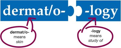
Dermatology is the medical specialty focused on the study, diagnosis, and treatment of the integumentary system, which includes the skin, hair, nails, and subcutaneous tissue. The term 'dermatology' is derived from the Greek roots dermat/o- (meaning skin) and -logy (meaning study of).
Integumentary System: Structure and Function
Overview of the Integumentary System
The integumentary system covers the entire surface of the body and serves as the first line of defense against environmental hazards. It consists of the skin (the largest organ of the body), nails, and subcutaneous tissue.
Layers and Components of the Skin
The skin is composed of two primary layers: the epidermis (outermost layer) and the dermis (deeper layer). Beneath the dermis lies the subcutaneous tissue, which is primarily composed of adipose tissue. The skin also contains hair follicles, sebaceous (oil) glands, and sudoriferous (sweat) glands.
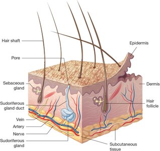
Anatomy of the Epidermis and Dermis
Epidermis
The epidermis is the thin, outermost layer of the skin, made up of epithelial tissue. It consists of two main sublayers:
Squamous layer: The upper part, containing both living and dead cells. Surface cells are dead, filled with keratin, and provide a protective barrier.
Basal layer: The deeper part, made of living cells that continuously divide and move upward. The epidermis contains no blood vessels and relies on the dermis for nutrients.
Melanocytes: Specialized cells in the basal layer that produce melanin, the pigment responsible for skin color and protection against UV radiation.
Dermis
The dermis lies beneath the epidermis and is composed of connective tissue, including collagen (providing strength) and elastin (providing elasticity). It contains blood vessels, nerves, hair follicles, sebaceous glands, and sweat glands.

Common Integumentary Diseases and Conditions
General Skin Conditions
Dermatitis: Inflammation or infection of the skin.
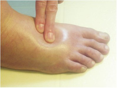
Edema: Swelling caused by excess fluid in the dermis or subcutaneous tissue.
Hemorrhage: Bleeding into the skin due to blood vessel injury.
Lesion: Any observable abnormality or damage to the skin.
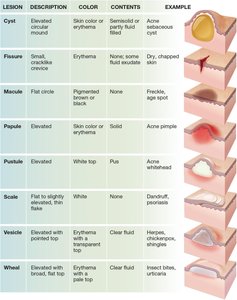
Types of Skin Lesions
Skin lesions are classified by their appearance, color, and contents. Common types include cysts, fissures, macules, papules, pustules, scales, vesicles, and wheals.
Lesion | Description | Color | Contents | Example |
|---|---|---|---|---|
Cyst | Elevated, circular mound | Skin color or erythema | Semisolid or partly fluid-filled | Acne sebaceous cyst |
Fissure | Small, cracklike crevice | Erythema | None, some fluid exudate | Dry, chapped skin |
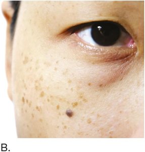
Macule | Flat circle | Pigmented brown or black | None | Freckle, age spot |
Papule | Elevated | Skin color or erythema | Solid | Acne pimple |
Pustule | Elevated | White top | Pus | Acne whitehead |
Scale | Flat to slightly elevated, thin flake | White | None | Dandruff, psoriasis |
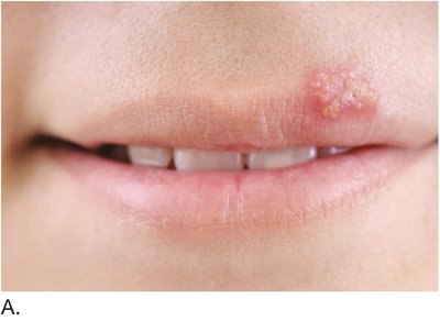
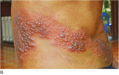
Vesicle | Elevated with pointed top | Erythema with transparent top | Clear fluid | Herpes, chickenpox |
Wheal | Elevated with broad, flat top | Erythema with pale top | Clear fluid | Insect bites, urticaria |
Skin Color Disorders
Albinism: Genetic absence of melanin, resulting in very light skin, hair, and eyes.
Cyanosis: Bluish-purple discoloration due to low oxygen levels in the blood.
Jaundice: Yellowish discoloration due to elevated bilirubin levels.
Necrosis: Gray-to-black discoloration from tissue death.
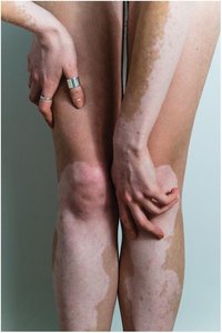
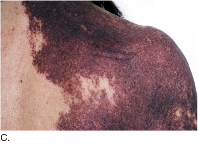
Vitiligo: Autoimmune destruction of melanocytes, causing white patches of skin.

Skin Injuries and Healing
Common Skin Injuries
Abrasion: Scraping injury that removes the epidermis.
Blister: Fluid-filled sac caused by friction separating the epidermis from the dermis.
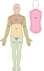
Burns: Classified by depth (superficial, partial-thickness, full-thickness) and extent (using the Rule of Nines).
Callus: Thickened pad of epidermis from repetitive rubbing.
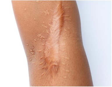
Cicatrix: Scar tissue formed during healing.
Keloid: Overgrown, firm scar due to excess collagen production.
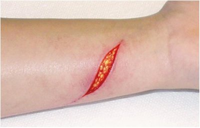
Laceration: Deep cut or tear in the skin.
Pressure injury: Ulcer from prolonged pressure, often in immobile patients.
Skin Infections and Infestations
Bacterial, Viral, and Fungal Infections
Abscess: Localized pus pocket, often around hair follicles (furuncle/carbuncle).
Cellulitis: Spreading infection of skin and subcutaneous tissue.
Herpes: Viral infection causing clusters of vesicles (oral, genital, shingles).
Tinea: Fungal infection (ringworm) affecting various body parts.
Verruca: Wart caused by human papillomavirus.
Parasitic Infestations
Pediculosis: Infestation with lice and their eggs (nits).
Scabies: Infestation with mites that burrow under the skin, causing intense itching.
Allergic and Autoimmune Skin Disorders
Allergic Reactions
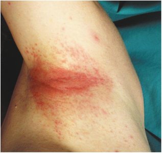
Contact dermatitis: Local reaction to allergens or irritants.
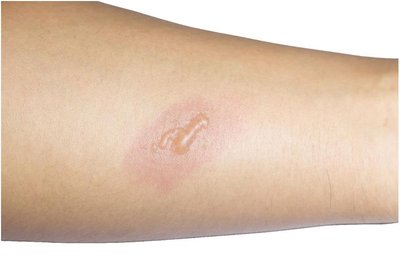
Urticaria (hives): Raised, red, itchy areas due to allergic triggers.
Autoimmune Disorders
Psoriasis: Excessive production of abnormal epidermal cells, causing red, scaly plaques.
Scleroderma: Hardening of skin and internal organs due to abnormal collagen deposits.
Systemic lupus erythematosus (SLE): Deterioration of collagen in skin and connective tissues, often with a butterfly rash.
Benign and Malignant Neoplasms of the Skin
Benign Neoplasms
Actinic keratosis: Rough, sun-exposed lesion that may become cancerous.
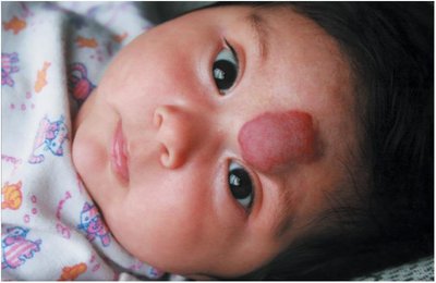
Hemangioma: Benign mass of dilated blood vessels, present at birth.
Lipoma: Benign fatty tumor in the subcutaneous layer.
Nevus (mole): Pigmented skin lesion, present at birth or acquired.
Papilloma: Soft, flesh-colored growth on a stalk.
Senile lentigo: Age spots from sun exposure.
Syndactyly: Fusion of skin and tissues between fingers or toes.
Malignant Neoplasms
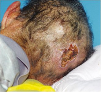
Basal cell carcinoma: Most common, slow-growing skin cancer, rarely metastasizes.
Squamous cell carcinoma: Begins in the squamous layer, slow-growing red bump or ulcer.
Malignant melanoma: Aggressive cancer of melanocytes, can metastasize rapidly. Diagnosed using the ABCDE criteria (Asymmetry, Border, Color, Diameter, Evolving).
Kaposi sarcoma: Cancer of connective tissue, often seen in immunocompromised patients.
Anatomy and Diseases of Skin Appendages
Hair
Hair covers most of the body, with color determined by melanocytes. Hair forms in follicles in the dermis and is composed of keratin. Piloerection (goosebumps) occurs when tiny muscles contract in response to cold or emotion.
Diseases of Hair
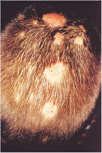
Alopecia: Hair loss due to genetics, disease, or hormonal changes.
Hirsutism: Excessive hair growth in females, often due to hormonal imbalance.
Nails
Nails consist of the nail plate, nail bed, lunula, cuticle, and nail root. They protect the distal phalanges and aid in fine manipulation.
Diseases of Nails
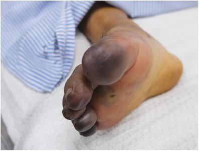
Clubbing and cyanosis: Downward curvature and bluish color due to chronic hypoxia.
Onychomycosis: Fungal infection causing thickened, discolored nails.
Subcutaneous Tissue
The subcutaneous layer (sub Q) is composed of loose connective and adipose tissue. It cushions, insulates, and protects internal organs. Thickness varies with fat storage.
Physiology of the Integumentary System
Protection
The skin acts as a barrier against injury, infection, and dehydration.
Keratin and acidic pH discourage microorganism growth.
Sweat and sebum contain antimicrobial substances.
Repair
Basal epidermal cells migrate to cover wounds.
Deep wounds form clots and scabs, followed by new cell growth.
Sensation
Sensory receptors in the dermis detect touch, pressure, pain, and temperature.
Nerve signals are transmitted to the spinal cord and brain for interpretation.
Vitamin D Synthesis
UV rays convert epidermal cholesterol to vitamin D, essential for calcium absorption and bone health.
Sun exposure requirements vary by skin tone.
Thermoregulation
Subcutaneous fat conserves heat; piloerector muscles generate heat.
Sweat glands and dermal blood vessels help cool the body.
Dermatomes
A dermatome is a specific area of skin supplied by sensory fibers from a single spinal nerve. Dermatomes are named according to the spinal cord level (C, T, L, S).
Laboratory and Diagnostic Procedures
Allergy skin testing: Identifies allergens causing reactions.
Culture and sensitivity (C&S): Identifies bacteria and appropriate antibiotics.
RAST: Measures IgE levels in response to allergens.
Skin scraping and Tzanck test: Diagnose fungal and viral infections.
Wood lamp: Uses UV light to detect fungal infections and pigment disorders.
Medical and Surgical Procedures
Botox and collagen injections: Reduce wrinkles and scars.
Cryosurgery: Freezes and destroys lesions.
Curettage and debridement: Remove superficial or necrotic tissue.
Electrosurgery: Uses electrical current to remove lesions.
Incision and drainage (I&D): Removes pus from abscesses.
Laser surgery: Removes birthmarks, tattoos, and unwanted hair.
Skin grafting and dermatoplasty: Replace or repair damaged skin.
Liposuction: Removes excess fat from subcutaneous tissue.
Mohs surgery: Removes skin cancer layer by layer.
Rhytidectomy: Facelift to remove wrinkles.
Pharmacology: Drugs for Integumentary Disorders
Anesthetic drugs: Numb the skin for procedures.
Antibiotics: Treat bacterial infections.
Antifungals: Treat fungal infections.
Antihistamines: Reduce allergic symptoms.
Antivirals: Treat herpes infections.
Corticosteroids: Reduce inflammation.
Alopecia drugs: Promote hair growth.
Drugs for infestations: Treat lice and scabies.
Psoriasis drugs: Slow cell production and reduce scaling.
Vitamin A-type drugs: Treat acne by increasing cell turnover.
Routes of Administration
Topical: Applied directly to the skin for local effect.
Transdermal: Patch delivers drug systemically through the skin.
Intradermal: Injection just under the epidermis (e.g., allergy testing).
Hypodermic: Injection into the subcutaneous tissue.
Common Abbreviations in Dermatology
Abbreviation | Definition |
|---|---|
B x | biopsy |
C & S | culture and sensitivity |
Derm | dermatology |
HSV | herpes simplex virus |
I & D | incision and drainage |
IgE | immunoglobulin E |
PDT | photodynamic therapy |
RAST | radioallergosorbent test |
SLE | systemic lupus erythematosus |
SQ, sub Q, subcu | subcutaneous |
