 Back
BackIntegumentary System: Medical Terminology and Anatomy Study Guide
Study Guide - Smart Notes
 Tailored notes based on your materials, expanded with key definitions, examples, and context.
Integumentary System
Overview and Functions
The integumentary system is a complex organ system that includes the skin, hair, nails, sebaceous glands, and sweat glands. It serves as the body's primary interface with the external environment and fulfills several essential functions:
Protection: Acts as a two-way barrier, preventing entry of pathogens and loss of body fluids.
Temperature Regulation: Maintains homeostasis by controlling heat loss and retention.
Sensory Reception: Contains receptors for touch, pain, pressure, and temperature.
Secretion: Produces sweat and sebum, which help cool and lubricate the skin.
Organs of the integumentary system: Skin, hair, nails, sebaceous glands, sweat glands.
Anatomy and Physiology of the Skin
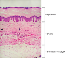
The skin is the largest organ in the human body, weighing over 20 pounds in adults. It is also known as the integument or cutaneous membrane. The skin is composed of three main layers, each with distinct functions and structures.
Epidermis: The thin, outer membrane layer made of stratified squamous epithelium. It contains no blood vessels or connective tissue.
Dermis: The middle, fibrous connective tissue layer, rich in blood supply and sensory receptors.
Subcutaneous Layer (Hypodermis): A layer of fatty tissue beneath the skin, providing insulation and protection.
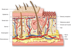
Layers of the Skin
Each layer of the skin has specialized functions:
Epidermis:
Composed of flat, scale-like cells arranged in strata.
The basal layer is the deepest part, where new cells are produced and pushed upward.
Cells fill with keratin as they move up, forming a waterproof barrier.
Contains melanocytes that produce melanin, the pigment responsible for skin color and protection against UV radiation.
Dermis:
Located between the epidermis and subcutaneous layer.
Made of connective tissue and collagen fibers, providing strength and elasticity.
Houses hair follicles, sweat and sebaceous glands, blood and lymph vessels, sensory receptors, and muscle fibers.
Dermal ridges form unique fingerprints.
Subcutaneous Layer:
Composed of fat cells (lipocytes).
Acts as insulation and protects deeper tissues from temperature extremes.
Accessory Organs of the Skin
The skin contains several accessory organs that contribute to its functions:
Hair:
Consists of the follicle, root, shaft, and arrector pili muscle.
Hair color is determined by melanin.
Arrector pili muscle contracts to make hair stand up (goosebumps).
Nails:
Main part is the nail body (keratin plate).
Connected to underlying tissue by the nail bed.
Lunula is the crescent-shaped area at the base.
Nails grow from the root, covered by the cuticle; the free edge is trimmed.
Sebaceous Glands:
Release sebum into hair follicles.
Sebum lubricates hair and skin, preventing dryness and cracking.
Increased secretion during adolescence can cause acne; decreased secretion with age leads to dry skin and wrinkles.
Sweat Glands (Sudoriferous Glands):
Coiled glands in the dermis.
Sweat travels to the surface via ducts and pores, cooling the body as it evaporates.
Most sweat is colorless and odorless; apocrine gland sweat produces body odor.
Key Medical Terminology
Understanding medical terminology is essential for describing the anatomy, physiology, and pathology of the integumentary system. Key elements include:
Combining Forms: Word roots combined with vowels to facilitate pronunciation (e.g., derm/o for skin).
Prefixes: Added to the beginning of terms to modify meaning (e.g., epi- meaning "upon").
Suffixes: Added to the end of terms to indicate procedures, conditions, or diseases (e.g., -itis meaning inflammation).
Examples and Applications
Keratinization: The process by which epidermal cells fill with keratin and die, forming a protective layer.
Melanin: Protects against UV damage and determines skin color; disorders include vitiligo and melanoma.
Sebum: Essential for maintaining skin moisture; overproduction leads to acne.
Sweat: Regulates body temperature; excessive sweating is called hyperhidrosis.
Summary Table: Layers of the Skin
Layer | Main Components | Functions |
|---|---|---|
Epidermis | Stratified squamous epithelium, melanocytes | Protection, waterproof barrier, pigmentation |
Dermis | Connective tissue, collagen, glands, follicles | Strength, elasticity, sensory reception, nourishment |
Subcutaneous Layer | Lipocytes (fat cells) | Insulation, energy storage, protection |
Additional info:
Medical terminology for the integumentary system is foundational for describing skin diseases, diagnostic procedures, and treatments.
Understanding the structure and function of the skin is essential for recognizing pathological changes and applying appropriate medical interventions.
