 Back
BackIntegumentary System: Structure, Pathology, and Clinical Procedures
Study Guide - Smart Notes
 Tailored notes based on your materials, expanded with key definitions, examples, and context.
Integumentary System
Overview and Functions
The integumentary system, primarily composed of the skin, is the body's largest organ system and serves multiple essential functions. It acts as a protective barrier, regulates body temperature, provides sensory information, eliminates waste, and initiates vitamin D synthesis.
Protection: Shields underlying tissues from mechanical damage, pathogens, and UV radiation. Melanin provides pigmentation and UV protection.
Temperature Regulation: Sweat glands and blood vessels help maintain homeostasis.
Sensory Reception: Specialized nerve endings detect touch, pressure, pain, and temperature.
Excretion: Perspiration removes metabolic waste products.
Vitamin D Synthesis: The skin initiates the production of vitamin D upon exposure to sunlight.
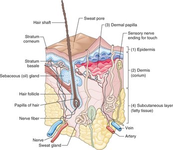
Skin Structure
The skin consists of three main layers, each with distinct structures and functions. Accessory structures include hair, nails, and glands.
Epidermis: The outermost layer, primarily protective, composed of stratified squamous epithelium.
Dermis: The middle, thicker layer containing connective tissue, blood vessels, nerves, hair follicles, and glands.
Subcutaneous Layer (Hypodermis): Composed mainly of loose connective and adipose tissue, providing insulation and cushioning.
Dermatology is the medical specialty focused on the study and treatment of skin disorders.
Accessory Structures
Hair: Provides protection and sensory input.
Nails: Protect the distal phalanges and enhance fine touch.
Glands: Include sebaceous (oil) and sweat glands, involved in lubrication and thermoregulation.
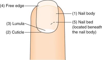
Nail Structure
Nails are composed of keratinized cells and have several distinct parts:
Nail Body: Visible, main part of the nail.
Cuticle: Protective fold of skin at the base of the nail.
Lunula: Crescent-shaped, whitish area at the nail base.
Nail Bed: Skin beneath the nail body.
Free Edge: The part of the nail that extends beyond the finger or toe.
Skin Lesions
Types of Lesions
Skin lesions are abnormal changes in skin structure due to disease or injury. They are classified by appearance and content:
Abrasion: Superficial scraping or rubbing of the skin (e.g., carpet burn).
Abscess: Localized collection of pus due to infection (e.g., pustule).
Blister (Vesicle): Small, fluid-filled lesion; a large blister is called a bulla.
Carbuncle: Deep skin infection with pus, often involving hair follicles.
Comedo: Acne lesion; closed (whitehead) or open (blackhead).
Cyst: Closed sac containing fluid, semifluid, or solid material.
Fissure: Crack-like sore or groove (e.g., anal fissure).
Fistula: Abnormal passage between organs or to the body surface.
Hives (Wheals): Elevated, pale-centered lesions (e.g., mosquito bite).
Laceration: Tear in the skin.
Macule: Flat, discolored spot (e.g., freckles, bruises).
Nodule: Small, raised swelling.
Papule: Small, solid elevation (e.g., pimple).
Polyp: Stalk-like growth from mucous membrane (e.g., nasal polyp).
Pustule: Small, pus-filled elevation (e.g., small abscess).
Scales: Flakes of hardened epithelium shed from the epidermis.
Ulcer: Open sore with inflammation (e.g., decubitus ulcer).
Wheal: Slightly elevated, pale-centered lesion (e.g., hives).
Combined Lesions: A rash with both macules and papules is termed maculopapular.
Pathological Conditions of the Skin
Common Disorders
Acne Vulgaris: Inflammatory disorder with papules, pustules, and comedos, commonly on the face, chest, and back.
Albinism: Inherited absence of pigment in skin, hair, and eyes.
Burns: Tissue injury classified by depth:
First-degree: Superficial (e.g., sunburn)
Second-degree: Partial-thickness (e.g., scalds)
Third-degree: Full-thickness (e.g., fire burns)
Callus: Thickened epidermis from friction or pressure.
Skin Cancers
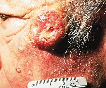
Basal Cell Carcinoma: Most common malignant epithelial tumor; slow-growing, often on sun-exposed skin. Begins as a nodule with central ulceration.
Squamous Cell Carcinoma: Malignancy of squamous cells; faster growing, higher risk of metastasis. Common on sun-exposed areas (nose, forehead, ears, hands, lower lip).
Malignant Melanoma: Tumor from melanocytes, often in pigmented skin. Noted for asymmetry, irregular borders, color variation, and diameter (>6mm). ABCDs help in recognition: Asymmetry, Borders, Color, Diameter.
Inflammatory and Infectious Conditions
Dermatitis: Inflammation of the skin, acute or chronic, contact or seborrheic.
Eczema: Chronic inflammatory condition with erythema, papules, vesicles, pustules, scales, and itching.
Exanthematous Viral Diseases: Viral rashes (e.g., rubella, roseola, rubeola, erythema infectiosum).
Gangrene: Tissue death from loss of blood supply and infection; can be dry or moist.
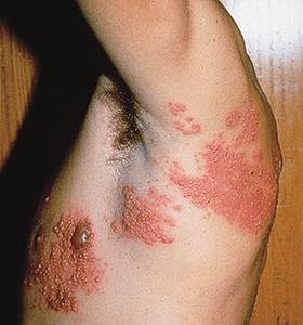
Herpes Zoster (Shingles): Acute viral infection with painful, vesicular eruptions along nerve pathways, common in adults over 50.
Hyperkeratosis: Overgrowth of the epidermis, seen in psoriasis, calluses, and corns.
Impetigo: Contagious bacterial infection with vesicles and pustules, often caused by staphylococcus or streptococcus.
Kaposi’s Sarcoma: Rare malignant lesions, often in AIDS patients, starting as purple-brown nodules.
Keloid: Enlarged, elevated scar from excess collagen.
Keratosis: Thickening and overgrowth of the cornified epithelium (e.g., seborrheic or actinic keratosis).
Leukoplakia: White, thickened patches on mucous membranes.
Nevus: Accumulation of melanocytes (mole).
Onychocryptosis: Ingrown nail, usually the big toe.
Onychomycosis: Fungal infection of the nails.
Pediculosis: Lice infestation (head, body, eyelashes, pubic hair).
Pemphigus: Rare disorder with widespread blisters.
Pilonidal Cyst: Sacrococcygeal cyst, sometimes present at birth.
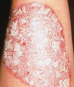
Psoriasis: Chronic, noninfectious disorder with silvery scales over red plaques, causing itching.
Rosacea: Chronic inflammation with redness, mainly on the face.
Scabies: Parasitic infestation by the human itch mite, causing rash and pruritus.
Scleroderma: Gradual thickening and hardening of the skin.
Systemic Lupus Erythematosus: Multisystem inflammatory disease with characteristic butterfly rash.
Tinea: Fungal infection (ringworm) affecting scalp, body, groin, or feet.
Verruca (Wart): Benign, elevated lesion from HPV infection; types include common, plantar, venereal, and seborrheic warts.
Diagnostic Techniques, Treatments, and Procedures
Common Diagnostic and Therapeutic Procedures
Allergy Testing: Identifies allergens via skin exposure (intradermal, patch, scratch tests) or blood tests (e.g., ImmunoCAP®).
Cautery: Burns and scars tissue using heat or chemicals.
Cryosurgery: Destroys tissue by freezing.
Curettage and Electrodesiccation: Scrapes away abnormal tissue, then destroys the base with electrical current.
Debridement: Removes debris, foreign objects, and necrotic tissue to prevent infection and promote healing.
Dermabrasion: Removes superficial skin layers to treat scars or tattoos.
Dermatoplasty: Skin transplantation for damaged areas.
Electrodesiccation: Burns and destroys tissue with an electrical spark (fulguration).
Electrosurgery: Removes or destroys tissue using electrical current.
Escharotomy: Incision into necrotic tissue from severe burns.
Liposuction: Removes fat via suction to alter body contours.
Skin Biopsy: Removes a small tissue sample for microscopic examination.
Skin Graft: Transplants skin to areas unable to regenerate.
Wood’s Lamp: Uses ultraviolet light to detect fungal infections of the skin and scalp.
Note: Debridement is for removing necrotic tissue and debris, while dermabrasion is for removing superficial skin layers for cosmetic or therapeutic reasons.
