 Back
BackIntroduction to the Respiratory System: Medical Terminology and Disorders
Study Guide - Smart Notes
 Tailored notes based on your materials, expanded with key definitions, examples, and context.
Introduction to the Respiratory System
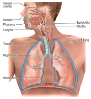
Overview of the Respiratory Tract
The respiratory system is responsible for the exchange of gases essential for life. It consists of a series of structures that conduct air into and out of the lungs, where gas exchange occurs.
Nose: Entry point for air.
Pharynx: Muscular tube connecting the nasal cavity to the larynx.
Larynx: Voice box; passageway for air and sound production.
Trachea: Windpipe; conducts air to the bronchi.
Bronchi and Bronchioles: Branching airways leading to the alveoli.
Alveoli: Air sacs where gas exchange occurs.
Lungs: Main organs of respiration.
Processes of Respiration
Ventilation: Movement of air in (inspiration) and out (expiration) of the lungs.
External Respiration: Exchange of gases between alveoli and blood.
Transport: Movement of gases in the blood between lungs and tissues.
Internal Respiration: Exchange of gases between blood and body tissues.
Functions of the Respiratory System:
Gas exchange (O2 and CO2)
Regulation of blood pH
Protection (cilia, mucus, tonsils)
Voice production (vocal cords)
Olfaction (smell receptors)
Medical Terminology: Word Analysis
Root Elements and Terms
Pulmon-: Lung (e.g., pulmonary)
Spir-: To breathe (e.g., spirometry)
Upper Respiratory Tract
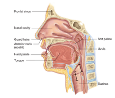
Structure and Function of the Nose
The nose serves as the main passageway for air entering the respiratory system. It filters, moistens, and warms incoming air and houses olfactory receptors for smell.
Nares: External openings of the nose.
Septum: Divides the nasal cavity into right and left halves.
Palate: Hard and soft palate separate the nasal and oral cavities.
Conchae and Meatus: Increase surface area for air filtration and humidification.
Disorders of the Nose
Upper Respiratory Infection (URI): Common viral infection affecting the nose and throat.
Rhinitis (Coryza): Acute inflammation of the nasal mucosa, usually viral.
Allergic Rhinitis: Allergic reaction causing clear, watery nasal discharge.
Sinusitis: Inflammation of the paranasal sinuses.
Deviated Nasal Septum: Displacement of the septum to one side.
Epistaxis: Nosebleed, often from the nasal septum.
Nasal Polyps: Benign mucosal growths in the nasal cavity or sinuses.
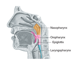
Pharynx: Structure and Function
The pharynx, or throat, is a muscular tube that serves as a passageway for both air and food. It is divided into three regions:
Nasopharynx: Posterior to the nasal cavity.
Oropharynx: Posterior to the oral cavity.
Laryngopharynx: Posterior to the larynx; leads to the esophagus and larynx.
Tonsils: Lymphatic tissue (palatine, pharyngeal/adenoid, and lingual) that helps protect against pathogens.
Uvula: Extension of the soft palate.
Disorders of the Pharynx
Snoring: Sound produced by vibration of soft tissues during sleep.
Obstructive Sleep Apnea (OSA): Repeated episodes of airway obstruction during sleep, causing hypoxia.
Apnea: Complete cessation of breathing.
Hypoxia: Low oxygen levels in tissues.
Pharyngitis: Inflammation of the pharynx.
Tonsillitis: Inflammation of the tonsils, often viral.
Nasopharyngeal Carcinoma: Cancer of the nasopharynx.
Medical Terminology: Adjective Forms
Pharynx → Pharyngeal: Pertaining to the pharynx
Nasopharynx → Nasopharyngeal: Pertaining to the nasopharynx
Oropharynx → Oropharyngeal: Pertaining to the oropharynx
Laryngopharynx → Laryngopharyngeal: Pertaining to the laryngopharynx
Larynx: Structure and Function
The larynx, or voice box, is a cartilaginous structure involved in sound production and maintaining an open airway.
Unpaired Cartilages: Thyroid (Adam's apple), cricoid, epiglottis
Paired Cartilages: Arytenoid, corniculate, cuneiform
Vocal Cords: True vocal folds for sound production
Vestibular Folds: False vocal cords
Major Roles:
Maintaining an open airway
Producing sound
Disorders of the Larynx
Laryngitis: Inflammation of the laryngeal mucosa
Epiglottitis: Inflammation of the epiglottis, common in young children
Laryngotracheobronchitis (Croup): Viral inflammation and obstruction of the upper airway in young children
Papilloma/Polyps: Benign tumors due to overuse or irritation
Lower Respiratory Tract
Structure and Function
Trachea: Windpipe; supported by C-shaped cartilage rings
Lungs: Right lung (3 lobes), left lung (2 lobes)
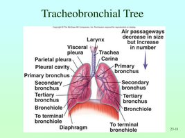
Tracheobronchial Tree: Branching system of airways
Bronchioles and Alveoli: Smallest airways and air sacs for gas exchange
Pleurae: Double-layered serous membrane covering the lungs
Bronchioles, Alveoli, and the Respiratory Membrane
Bronchioles: Smallest conducting airways
Terminal Bronchioles: End of conducting zone
Alveoli: Site of gas exchange
Respiratory Membrane: Thin barrier (0.5 µm) for gas exchange; consists of alveolar epithelium, fused basement membranes, and capillary endothelium
Function: Oxygen diffuses from alveoli to blood; carbon dioxide diffuses from blood to alveoli.
Surfactant
Definition: Lipoprotein secreted by type II alveolar cells
Function: Reduces surface tension, prevents alveolar collapse, aids lung inflation
Clinical Relevance: Deficiency in premature infants causes Infant Respiratory Distress Syndrome (IRDS); damage can lead to breathing difficulties
Tracheobronchial Tree Pathway
Nose → Pharynx → Larynx → Trachea → Primary Bronchi → Secondary Bronchi → Tertiary Bronchi → Bronchioles → Alveoli
Each alveolus is surrounded by a pulmonary capillary network for gas exchange.
Lungs and Pleura
Right Lung: Three lobes
Left Lung: Two lobes
Pleura: Double-layered membrane (visceral and parietal)
Pleural Cavity: Space between pleural layers, filled with pleural fluid
Disorders of the Respiratory System
Common Symptoms and Signs
Cough: Reflex to clear irritants; hemoptysis is coughing up blood
Dyspnea: Shortness of breath
Cyanosis: Bluish discoloration due to unoxygenated hemoglobin
Changes in Breathing Rate:
Eupnea: Normal breathing (10–15 breaths/min in adults)
Tachypnea: Rapid breathing
Hyperpnea: Deep, rapid breathing
Bradypnea: Slow breathing
Major Respiratory Disorders
Asthma: Recurrent bronchial obstruction, bronchoconstriction, mucus hypersecretion, airway inflammation
Hypercapnia: Excess carbon dioxide in the blood
Chronic Bronchitis: Long-term airway inflammation, excess mucus, cilia destruction; often due to smoking
Acute Bronchitis: Short-term inflammation, usually viral or bacterial; resolves without lasting damage
COPD (Chronic Obstructive Pulmonary Disease): Group of diseases (mainly chronic bronchitis and emphysema) causing long-term airflow limitation
Emphysema: Destruction of alveolar walls, reducing gas exchange surface area
Cystic Fibrosis (CF): Genetic disorder causing thick, sticky mucus in lungs and digestive tract
Infant Respiratory Distress Syndrome (IRDS): Premature infants lack surfactant, leading to breathing difficulties
Pleurisy (Pleuritis): Inflammation of the pleura, often due to infection or other diseases
Pneumothorax: Air in the pleural space causing lung collapse
Pneumonia: Infection of the lungs, alveoli fill with fluid or pus, impairing gas exchange
Summary Table: Key Respiratory Disorders
Disorder | Main Feature | Cause |
|---|---|---|
Asthma | Bronchial constriction, mucus, inflammation | Allergy, irritants |
Chronic Bronchitis | Chronic airway inflammation, mucus | Smoking, infection |
Emphysema | Destruction of alveolar walls | Smoking, genetic |
Cystic Fibrosis | Thick mucus in lungs/digestive tract | Genetic |
IRDS | Lack of surfactant in infants | Prematurity |
Pleurisy | Pleural inflammation | Infection, autoimmune |
Pneumothorax | Air in pleural space, lung collapse | Trauma, disease |
Pneumonia | Alveoli filled with fluid/pus | Infection |
Additional info: This guide expands on the provided notes with definitions, clinical context, and a summary table for clarity and exam preparation.
