 Back
BackMedical Terminology and Anatomy of the Gastrointestinal System
Study Guide - Smart Notes
 Tailored notes based on your materials, expanded with key definitions, examples, and context.
Gastroenterology: Medical Terminology
Word Roots and Structure
Medical terminology often uses word roots, prefixes, and suffixes to describe anatomical structures and diseases. Understanding these components is essential for proficiency in medical language.
gastr/o-: means stomach
enter/o-: means intestine
-logy: means study of
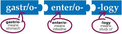
Example: Gastroenterology is the study of the stomach and intestines.
Anatomy of the Gastrointestinal (GI) System
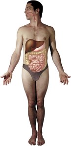
Overview of GI System
The gastrointestinal system consists of organs and glands connected in a pathway. Food enters the body, is digested, nutrients are absorbed into the blood, and undigested waste is eliminated.
Oral Cavity and Pharynx
The oral cavity contains the teeth, gums, tongue, and the hard and soft palates. Food passes from the oral cavity into the pharynx (throat) and then the esophagus.
Teeth: Responsible for mastication (chewing).
Gums: Support the teeth.
Tongue: Perceives taste and aids in swallowing.
Hard and Soft Palate: Form the roof of the mouth.

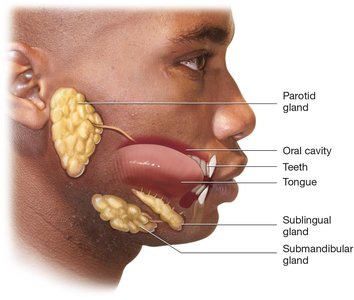
Salivary Glands
Salivary glands produce saliva, which begins the process of digestion and lubricates food for swallowing. The major salivary glands are:
Parotid glands: Located anterior to the ear.
Sublingual glands: Located under the tongue.
Submandibular glands: Located inferior to the mandible.
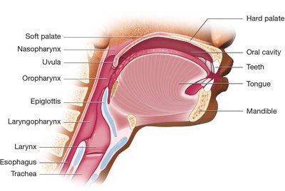
Pharynx and Larynx
The pharynx is a common passageway for air and food, divided into three parts: nasopharynx, oropharynx, and laryngopharynx. The larynx is just inferior to the pharynx and remains open during breathing and speaking, but closes during swallowing to prevent food from entering the trachea.
Epiglottis: Seals the opening to the larynx during swallowing.

Esophagus
The esophagus is a muscular tube connecting the pharynx to the stomach. Peristalsis moves food through the esophagus.
Upper esophagus: Located in the neck.
Lower esophagus: Located in the thoracic cavity.
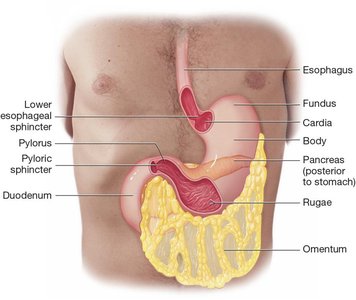
Stomach Anatomy
The stomach is divided into four parts: cardia, fundus, body, and pylorus. The omentum is a fatty membrane protecting abdominal organs. The stomach's mucosa forms rugae, which expand to accommodate food.
Cardia: Where the stomach joins the esophagus.
Fundus: Rounded upper portion.
Body: Large curved region.
Pylorus: Narrow last part joining the duodenum.
Rugae: Folds in the gastric mucosa.
Sphincters: Lower esophageal and pyloric sphincters regulate passage of food.
Diseases of the GI System
Oral Cavity and Salivary Gland Diseases
Glossitis: Infection or inflammation of the tongue.
Sialolithiasis: Presence of a stone in a salivary gland causing swelling.
Esophagus and Stomach Diseases
Dyspepsia: Temporary epigastric pain; indigestion.
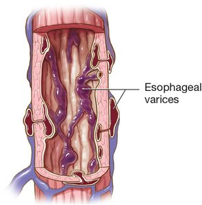
Esophageal varix: Swollen, protruding vein in the esophageal mucosa.
Gastritis: Acute or chronic inflammation of the stomach.
Gastroenteritis: Acute infection or inflammation of the stomach and intestines.
Gastroesophageal reflux disease (GERD): Chronic irritation due to reflux of stomach acid into the esophagus.
Peptic ulcer disease (PUD): Chronic irritation and erosion of the mucosa due to an ulcer.
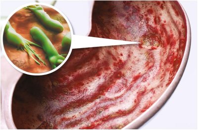
Stomach cancer: Cancerous tumor of the stomach, often caused by H. pylori.
Diagnostic and Surgical Procedures
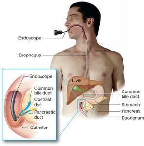
Endoscopy and Related Procedures
Endoscopy uses a flexible, fiberoptic scope to examine the GI system internally. Endoscopic retrograde cholangiopancreatography (ERCP) visualizes the bile and pancreatic ducts using contrast dye.
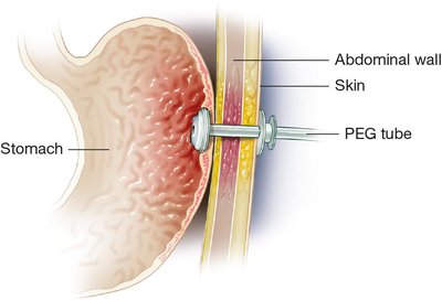
Nasogastric and PEG Tubes
Nasogastric (NG) tubes are inserted through the nose into the stomach for feeding or drainage. Percutaneous endoscopic gastrostomy (PEG) tubes are inserted through the abdominal wall for permanent feeding access.

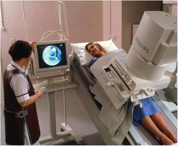
Radiologic Procedures
Upper gastrointestinal series (UGI) uses barium contrast to outline the esophagus, stomach, and duodenum. Oral cholecystography (OCG) uses iodinated contrast dye to visualize the gallbladder.
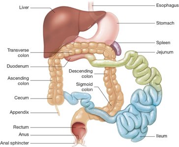
Anatomy: Small and Large Intestines
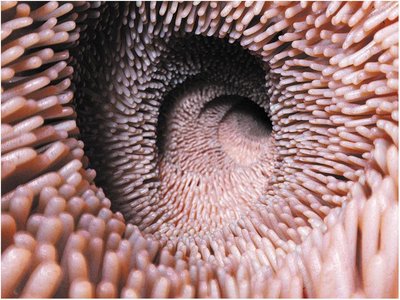
Small Intestine
The small intestine consists of the duodenum, jejunum, and ileum. It contains thousands of villi to maximize absorption of nutrients and water.
Duodenum: First segment, C-shaped, ~10 inches long.
Jejunum: Middle segment, ~8 feet long.
Ileum: Third segment, ~12 feet long.
Diseases of the Small Intestine
Ileus: Absence of normal peristalsis.
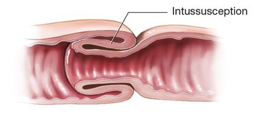
Intussusception: Telescoping of one intestinal segment inside another.
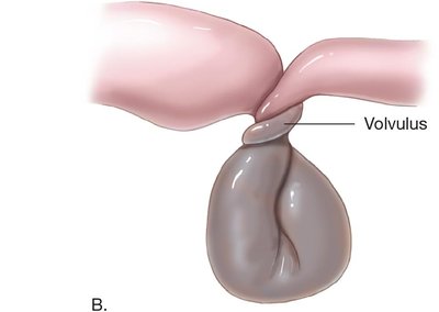
Volvulus: Twisting or rotating of the intestine.
Large Intestine
The large intestine runs from the small intestine to the outside surface of the body. It consists of the cecum, colon, and rectum. The colon is divided into ascending, transverse, descending, and sigmoid parts.
Diseases of the Cecum and Colon
Appendicitis: Inflammation and infection of the appendix.
Colon cancer: Cancerous tumor of the colon.
Diverticulum/Diverticulitis: Pouches in the colon wall that can become infected.
Gluten sensitivity enteropathy: Autoimmune reaction to gluten.
Inflammatory bowel disease (IBD): Chronic inflammation (Crohn disease, ulcerative colitis).
Irritable bowel syndrome (IBS): Functional disorder of the colon.
Polyposis: Numerous polyps in the colon.
Rectum and Anus
Anatomy and Diseases
Hemorrhoids: Swollen veins in the rectum.
Proctitis: Inflammation of the rectum and anus.
Rectocele: Hernia in the wall of the rectum.
Defecation and Feces
Constipation: Failure to have regular, soft bowel movements.
Diarrhea: Frequent, loose, watery feces.
Flatulence: Excessive gas in the stomach or intestines.
Hematochezia: Blood in the feces.
Steatorrhea: Feces containing undigested fats.
Abdominopelvic Cavity and Accessory Organs
Liver
The liver is the largest solid organ, producing bile and processing nutrients. Diseases include cirrhosis, hepatomegaly, hepatitis, jaundice, and liver cancer.
Gallbladder and Bile Ducts
The gallbladder stores and concentrates bile. Diseases include cholangitis, cholecystitis, cholelithiasis, and gallbladder cancer.
Pancreas
The pancreas secretes digestive enzymes into the duodenum. Diseases include pancreatic cancer and pancreatitis.
Physiology of Digestion
Mechanical Digestion
Mastication: Chewing by the teeth.
Deglutition: Swallowing food.
Peristalsis: Muscular contractions moving food.
Chemical Digestion
Amylase: Breaks down carbohydrates.
Pepsin: Breaks down proteins.
Gastrin: Stimulates release of hydrochloric acid and pepsinogen.
Hydrochloric acid: Breaks down food fibers.
Cholecystokinin: Stimulates gallbladder to release bile.
Bile: Breaks down fats.
Lactase: Breaks down milk sugars.
Absorption and Elimination
Absorption: Nutrients and water absorbed mainly in the small intestine.
Elimination: Removal of undigested waste as feces.
Laboratory Tests and Diagnostic Procedures
Albumin: Major protein in blood, produced by the liver.
Cologuard®: DNA screening test for cancer in stool.
Culture and sensitivity (C & S): Stool test for bacteria.
Fecal occult blood test: Detects hidden blood in feces.
Ova and parasites (O & P): Stool test for parasites.
Helicobacter pylori tests: Detects H. pylori infection.
Liver function tests (LFTs): Panel of blood tests for liver function.
Common Drugs for GI Diseases
Antacid drugs: Neutralize stomach acid.
Antibiotic drugs: Treat bacterial infections.
Antidiarrheal drugs: Slow peristalsis and increase water absorption.
Antiemetic drugs: Treat nausea and vomiting.
Antispasmodic drugs: Decrease peristalsis and spasms.
Bile acid drugs: Decrease cholesterol production for gallstones.
H2 blocker drugs: Block histamine receptors to reduce acid.
Laxative drugs: Treat constipation.
Proton pump inhibitor drugs: Block production of hydrochloric acid.
Abbreviation Summary
Abbreviation | Definition |
|---|---|
ABD | abdomen |
ALP | alkaline phosphatase |
ALT | alanine aminotransferase |
AST | aspartate aminotransferase |
BE | barium enema |
BM | bowel movement |
C&S | culture and sensitivity |
CAT, CT | computerized axial tomography |
CBD | common bile duct |
EGD | esophagogastroduodenoscopy |
ERCP | endoscopic retrograde cholangiopancreatography |
GERD | gastroesophageal reflux disease |
IBD | inflammatory bowel disease |
IBS | irritable bowel syndrome |
LFTs | liver function tests |
MRI | magnetic resonance imaging |
N&V | nausea and vomiting |
NG | nasogastric |
OCG | oral cholecystogram |
PUD | peptic ulcer disease |
RLQ | right lower quadrant |
RUQ | right upper quadrant |
UGI | upper gastrointestinal (series) |
