 Back
BackMedical Terminology and Anatomy of the Gastrointestinal System
Study Guide - Smart Notes
 Tailored notes based on your materials, expanded with key definitions, examples, and context.
Gastroenterology: Medical Terminology
Word Roots and Structure
Medical terminology often uses word roots, prefixes, and suffixes to describe anatomical structures and diseases. Understanding these components is essential for proficiency in medical language.
gastr/o-: means stomach
enter/o-: means intestine
-logy: means study of
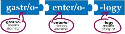
Example: Gastroenterology is the study of the stomach and intestines.
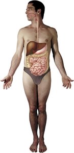
Anatomy of the Gastrointestinal (GI) System
Overview of GI System
The gastrointestinal system consists of organs and glands connected in a pathway. Its primary functions are digestion, absorption of nutrients, and elimination of waste.
Food enters the body, is digested, nutrients are absorbed into the blood, and undigested waste is eliminated.
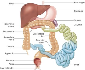
Main organs: oral cavity, pharynx, esophagus, stomach, small intestine, large intestine, rectum, and anus.
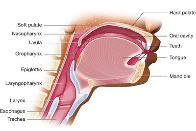
Oral Cavity and Pharynx
The oral cavity contains the teeth, gums, tongue, and the hard and soft palates. Food passes from the oral cavity into the pharynx (throat) and then the esophagus.
Teeth: Responsible for mastication (chewing).
Tongue: Contains taste receptors; aids in swallowing.
Hard and Soft Palate: Form the roof of the mouth.
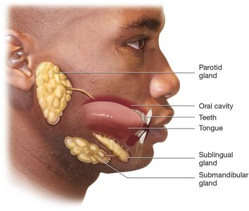
Salivary Glands
Salivary glands produce saliva, which begins the process of digestion and lubricates food for swallowing.
Parotid glands: Located anterior to the ear.
Sublingual glands: Located under the tongue.
Submandibular glands: Located inferior to the mandible.
Pharynx and Larynx
The pharynx is a common passageway for air and food, divided into nasopharynx, oropharynx, and laryngopharynx. The larynx is just inferior to the pharynx and remains open during breathing and speaking, but closes during swallowing to prevent food from entering the trachea.
Epiglottis: Seals the opening to the trachea during swallowing.

Esophagus
The esophagus is a muscular tube connecting the pharynx to the stomach. Peristalsis moves food through the esophagus.
Upper esophagus: Located in the neck.
Lower esophagus: Located in the thoracic cavity.

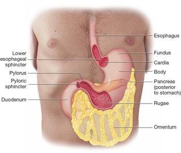
Stomach Anatomy
The stomach is divided into four parts: cardia, fundus, body, and pylorus. The omentum is a fatty membrane that protects abdominal organs.
Cardia: Where the stomach joins the esophagus.
Fundus: Rounded, upper portion.
Body: Large curved region.
Pylorus: Narrow last part joining the duodenum.
Rugae: Folds in the gastric mucosa that expand.
Sphincters: Lower esophageal and pyloric sphincters regulate passage of food.
Diseases of the Upper GI Tract
Common Diseases
Dyspepsia: Temporary epigastric pain; indigestion.
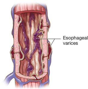
Esophageal varix: Swollen, protruding vein in the esophageal mucosa.
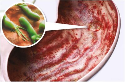
Gastritis: Acute or chronic inflammation of the stomach.
Gastroenteritis: Acute infection or inflammation of the stomach and intestines.
Diagnostic and Surgical Procedures
Endoscopy and Related Procedures
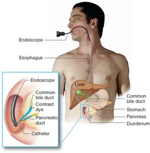
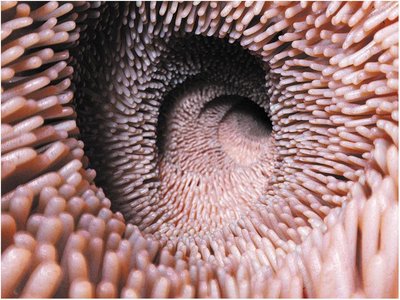
Endoscopy uses a flexible scope to examine the GI system internally. Endoscopic retrograde cholangiopancreatography (ERCP) visualizes the bile and pancreatic ducts using contrast dye.
Endoscopy: Internal examination of GI tract.
ERCP: Visualizes bile and pancreatic ducts.
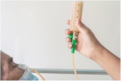
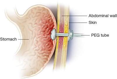
Nasogastric and PEG Tubes
Nasogastric (NG) tubes are used for temporary feeding or drainage. Percutaneous endoscopic gastrostomy (PEG) tubes are used for permanent feeding access.
NG tube: Inserted through nose into stomach.
PEG tube: Inserted through abdominal wall into stomach.
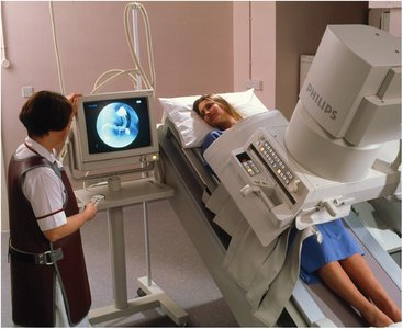
Radiologic Procedures
Upper gastrointestinal series (UGI) uses barium contrast to outline the GI tract for imaging.
Barium meal: Swallowed to coat GI tract for x-ray imaging.
Anatomy of the Lower GI Tract
Small and Large Intestines
The small intestine consists of the duodenum, jejunum, and ileum. The large intestine consists of the cecum, colon, rectum, and anus.
Small intestine: Absorbs nutrients and water; contains villi.
Large intestine: Absorbs water; forms and eliminates feces.
Diseases of the Small Intestine
Ileus: Absence of normal peristalsis.
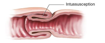
Intussusception: Telescoping of one intestinal segment inside another.
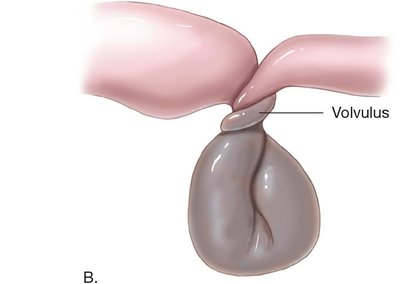
Volvulus: Twisting of the intestine.
Diseases of the Large Intestine
Common Diseases
Appendicitis: Inflammation of the appendix.
Colon cancer: Tumor of the colon.
Diverticulum: Pouch in the colon wall.
Inflammatory bowel disease (IBD): Chronic inflammation (Crohn disease, ulcerative colitis).
Irritable bowel syndrome (IBS): Functional disorder of the colon.
Polyposis: Numerous polyps in the colon.
Surgical Procedures of the Lower GI Tract
Colostomy and Stoma
Colostomy is the creation of an opening in the abdominal wall for feces to pass through. A stoma is the external opening.
Colostomy bag: Collects feces from the stoma.
Colonoscopy
Colonoscopy uses a colonoscope to examine the rectum and colon for abnormalities.
Radiologic Procedures: Barium Enema
Barium enema is a radiologic procedure that inserts barium contrast medium into the rectum to visualize the colon.
Diseases of the Abdominal Wall and Cavity
Hernia
A hernia is a defect in the muscle wall allowing the intestine and peritoneum to bulge through.
Sliding hernia: Intestine moves freely.
Strangulated hernia: Intestine is trapped and blood supply is cut off.
Omphalocele: Umbilical hernia present at birth.
Peritonitis
Peritonitis is infection and inflammation of the peritoneum, often due to perforation of the GI tract.
Anatomy and Diseases of Accessory Organs
Liver
The liver is the largest solid organ, producing bile and processing nutrients.
Cirrhosis: Chronic inflammation and degeneration.
Hepatomegaly: Enlargement of the liver.
Hepatitis: Infection and inflammation by hepatitis virus.
Jaundice: Yellow discoloration due to bilirubin.
Liver cancer: Tumor, often secondary from other sites.
Ascites: Fluid accumulation in the abdomen.
Gallbladder and Bile Ducts
The gallbladder stores and concentrates bile. Diseases include cholangitis, cholecystitis, cholelithiasis, and gallbladder cancer.
Gallstones: Can block bile ducts and cause complications.
Pancreas
The pancreas secretes digestive enzymes into the duodenum. Diseases include pancreatic cancer and pancreatitis.
Physiology of Digestion
Mechanical and Chemical Digestion
Digestion consists of mechanical (mastication, deglutition, peristalsis) and chemical (enzymes, acids, hormones) processes.
Mastication: Chewing by teeth.
Deglutition: Swallowing.
Peristalsis: Muscular contractions moving food.
Enzymes: Amylase, pepsin, gastrin, cholecystokinin, bile, lactase.
Absorption and Elimination
Absorption occurs mainly in the small intestine. Elimination removes undigested waste as feces.
Defecation: Process of eliminating feces.
Flatus: Gas expelled with feces.
Laboratory Tests and Drugs
Common Laboratory Tests
Albumin: Indicates liver function.
Cologuard®: Detects DNA of cancerous cells in stool.
C&S: Identifies bacteria in stool.
Fecal occult blood test: Detects hidden blood in feces.
Ova and parasites (O&P): Detects parasites in GI system.
Liver function tests (LFTs): Panel for liver health.
Common Drugs
Antacid: Neutralizes stomach acid.
Antibiotic: Treats bacterial infections.
Antidiarrheal: Treats diarrhea.
Antiemetic: Treats nausea and vomiting.
Antispasmodic: Treats spasms and cholecystitis.
Bile acid drug: Reduces gallstone formation.
H2 blocker: Treats GERD and peptic ulcer disease.
Laxative: Treats constipation.
Proton pump inhibitor: Blocks hydrochloric acid production.
Abbreviation Summary
Abbreviation | Definition |
|---|---|
ABD | abdomen |
ALP | alkaline phosphatase |
ALT | alanine aminotransferase |
AST | aspartate aminotransferase |
BE | barium enema |
BM | bowel movement |
C&S | culture and sensitivity |
CAT, CT | computerized axial tomography |
CBD | common bile duct |
EGD | esophagogastroduodenoscopy |
ERCP | endoscopic retrograde cholangiopancreatography |
GERD | gastroesophageal reflux disease |
IBD | inflammatory bowel disease |
IBS | irritable bowel syndrome |
LFTs | liver function tests |
NG | nasogastric |
PEG | percutaneous endoscopic gastrostomy |
PUD | peptic ulcer disease |
RLQ | right lower quadrant |
RUQ | right upper quadrant |
UGI | upper gastrointestinal (series) |
