 Back
BackMedical Terminology Study Guide: Gastrointestinal and Respiratory Systems
Study Guide - Smart Notes
 Tailored notes based on your materials, expanded with key definitions, examples, and context.
Gastroenterology & Gastrointestinal (GI) System
Overview of Gastroenterology
Gastroenterology is the medical specialty focused on the anatomy, physiology, and diseases of the gastrointestinal system. Gastroenterologists use diagnostic tests, medical and surgical procedures, and pharmacological treatments to manage GI diseases.
Gastroenterology: Study of the stomach and intestines.
GI System: Composed of organs and glands forming a pathway for digestion, absorption, and elimination.
Word Breakdown: Gastroenterology
Gastr/o-: Stomach
Enter/o- / Intestin/o-: Intestine
-logy: Study of
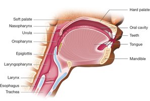
Upper GI System Anatomy
The upper GI system includes the oral cavity, pharynx, esophagus, and stomach. The oral cavity is the entry point for food and contains several important structures.
Oral Cavity: Contains teeth, gums, tongue, hard palate, and soft palate.
Pharynx: Common passageway for air and food.
Esophagus: Muscular tube connecting pharynx to stomach.
Peristalsis: Wave-like muscular contractions moving food through the digestive tract.
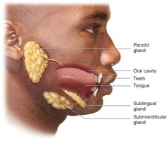
Salivary Glands
Salivary glands are triggered by sight, smell, and taste. They include:
Parotid Glands: Located anterior to the ear.
Sublingual Glands: Located under the tongue.
Submandibular Glands: Located inferior to the mandible.
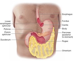
Stomach Anatomy
The stomach is an elongated sac with four main regions: cardia, fundus, body, and pylorus. It is lined by gastric mucosa and contains rugae for expansion.
Cardia: Junction with esophagus
Fundus: Rounded upper portion
Body: Large curved region
Pylorus: Narrow region joining duodenum
Lower Esophageal Sphincter: Controls entry from esophagus
Pyloric Sphincter: Controls exit to small intestine
Pancreatic Enzymes
Amylase: Breaks down carbohydrates
Lipase: Breaks down fats
Protein-Digesting Enzymes: Break down proteins
Small and Large Intestines
The small intestine is divided into three segments: duodenum, jejunum, and ileum. The large intestine includes the cecum, colon (ascending, transverse, descending, sigmoid), rectum, and anus.
Villi: Increase surface area for absorption in small intestine
Haustra: Pouches in colon for expansion
Ileocecal Valve: Prevents backflow from large to small intestine
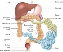
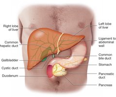
Accessory Organs
The liver, gallbladder, and pancreas are accessory organs supporting digestion.
Liver: Produces bile, processes nutrients
Gallbladder: Stores and concentrates bile
Pancreas: Secretes digestive enzymes and insulin
Digestion and Absorption
Digestion involves mechanical and chemical processes. Absorption occurs mainly in the ileum, with water absorption in the large intestine.
Mechanical Digestion: Mastication, deglutition, peristalsis
Chemical Digestion: Enzymatic breakdown of food
Absorption: Nutrients and water absorbed into blood
Common GI Diseases and Terminology
Medical terminology for GI diseases is based on word roots, prefixes, and suffixes. Examples include:
Anorexia: Decreased appetite
Dysphagia: Difficulty swallowing
Glossitis: Inflammation of the tongue
Gastritis: Inflammation of the stomach
GERD: Chronic reflux of stomach acid
Appendicitis: Inflammation of the appendix
Hepatitis: Inflammation of the liver
Laboratory and Diagnostic Procedures
Albumin: Liver function test
Cologuard: Stool DNA test for cancer
Fecal Occult Blood Test: Detects hidden blood in stool
Liver Function Tests (LFTs): Panel for liver health
Barium Enema: Radiologic imaging of colon
Gallbladder Ultrasound: Imaging for gallstones
Medical and Surgical Procedures
Enema: Relieves constipation
Appendectomy: Removal of appendix
Bariatric Surgery: Treats obesity
Colostomy: Creation of opening for fecal passage
Liver Transplantation: Replacement of diseased liver
Pulmonology: Respiratory System
Overview of Pulmonology
Pulmonology is the medical specialty focused on the anatomy, physiology, and diseases of the respiratory system. Pulmonologists diagnose and treat respiratory diseases using various tests and procedures.
Pulmon/o-: Lung
-logy: Study of
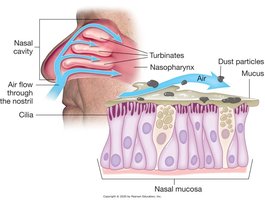
Anatomy: Upper Respiratory System
The upper respiratory system includes the nose, nasal cavity, and pharynx. The nasal cavity is lined with mucosa and contains turbinates to warm and moisten air.
Nose: Entry point for air, traps particles
Nasal Cavity: Divided by septum, contains turbinates
Nasal Mucosa: Produces mucus, humidifies air
Pharynx and Associated Structures
The pharynx is a common passage for air and food, divided into nasopharynx, oropharynx, and laryngopharynx.
Nasopharynx: Superior part
Oropharynx: Middle part
Laryngopharynx: Inferior part
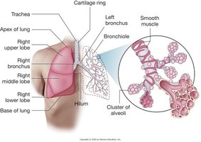
Anatomy: Lower Respiratory System
The lower respiratory system includes the larynx, trachea, bronchi, bronchioles, and lungs. The bronchial tree transports air to alveoli, where gas exchange occurs.
Larynx: Voice box, contains vocal cords
Trachea: Windpipe, supported by cartilage rings
Bronchi: Main airways entering lungs
Bronchioles: Smallest airways, end in alveoli
Alveoli: Site of gas exchange, secrete surfactant
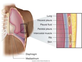
Lung Structure and Pleura
The lungs are divided into lobes and surrounded by pleura, a double-layered membrane that secretes pleural fluid for smooth movement.
Right Lung: Three lobes (RUL, RML, RLL)
Left Lung: Two lobes (LUL, LLL)
Pleura: Visceral and parietal layers, pleural fluid
Mediastinum: Central area containing heart, trachea, esophagus
Diaphragm: Muscle separating thoracic and abdominal cavities
Summary Table: GI and Respiratory System Structures
GI System Structure | Function |
|---|---|
Oral Cavity | Entry point for food, mechanical digestion |
Stomach | Mixes and digests food |
Small Intestine | Absorption of nutrients |
Large Intestine | Absorption of water, elimination |
Liver | Bile production, metabolism |
Gallbladder | Bile storage |
Pancreas | Enzyme and insulin secretion |
Respiratory System Structure | Function |
|---|---|
Nasal Cavity | Filters, warms, moistens air |
Pharynx | Passage for air and food |
Larynx | Voice production |
Trachea | Air passage to lungs |
Bronchi/Bronchioles | Air distribution |
Alveoli | Gas exchange |
Pleura | Reduces friction |
Additional info: Academic context and expanded explanations were added to ensure completeness and clarity for medical terminology students.
