 Back
BackMusculoskeletal System: Medical Terminology Study Guide
Study Guide - Smart Notes
 Tailored notes based on your materials, expanded with key definitions, examples, and context.
Musculoskeletal System Overview
Introduction
The musculoskeletal system is a fundamental component of human anatomy, providing structural support, movement, and protection for vital organs. It consists of bones, muscles, joints, and connective tissues, and is a major focus in medical terminology due to its complexity and clinical relevance.
Key Functions: Support, protection, movement, blood cell production, and mineral storage.
Major Organs: Bones, joints, muscles.
Skeletal System
Anatomy and Physiology of the Skeletal System
The skeletal system is composed of 206 bones, forming the internal framework of the body. Each bone is an organ with its own blood supply, nerves, and lymphatic vessels.
Functions: Supports the body, protects organs, provides muscle attachment points, produces blood cells (hematopoiesis), and stores minerals (e.g., calcium, phosphorus).
Bone Tissue: Also called osseous tissue, one of the hardest materials in the body.
Ossification: Gradual process where cartilage is replaced by bone cells (osteoblasts and osteocytes).
Remodeling: Old bone is broken down by osteoclasts and replaced; adult bones are completely renewed every 10 years.
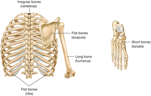
Classification of Bones by Shape
Bones are classified based on their shape and function.
Long Bones: e.g., femur, humerus
Short Bones: e.g., tarsals, carpals
Flat Bones: e.g., ribs, scapula
Irregular Bones: e.g., vertebrae
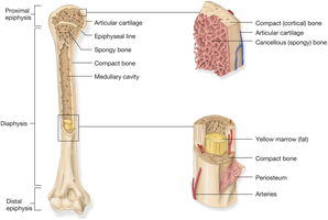
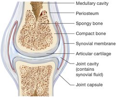
Structure of a Long Bone
Long bones have distinct anatomical features:
Diaphysis: Central shaft containing the medullary cavity (filled with yellow marrow).
Epiphysis: Wide ends of the bone, covered by articular cartilage.
Periosteum: Tough membrane covering the bone except at the joint surfaces.
Compact (Cortical) Bone: Hard exterior found in diaphysis and epiphysis.
Cancellous (Spongy) Bone: Interior with spaces containing red bone marrow.
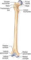
Bony Projections and Depressions
Bones have various projections and depressions, known as processes, which serve as points for muscle attachment or joint articulation.
Round, smooth processes: Allow for joint articulation.
Rough processes: Provide muscle attachment points.
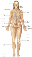
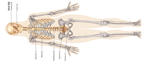
Divisions of the Skeleton
The skeleton is divided into two main parts:
Axial Skeleton: Head, neck, spine, chest, and trunk.
Appendicular Skeleton: Pectoral girdle, upper extremities, pelvic girdle, and lower extremities.
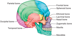
Skull Anatomy
The skull is divided into the cranium and facial bones.
Cranium: Frontal, parietal, temporal, ethmoid, sphenoid, occipital bones.
Facial Bones: Mandible, maxilla, zygomatic, vomer, palatine, nasal, lacrimal bones.
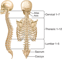
Vertebral Column
The vertebral column is divided into five regions:
Cervical: C1–C7
Thoracic: T1–T12
Lumbar: L1–L5
Sacrum: Fused vertebrae at the base
Coccyx: Tailbone
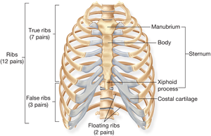
Rib Cage
The rib cage consists of 12 pairs of ribs:
True Ribs: First 7 pairs, attach directly to the sternum.
False Ribs: Next 3 pairs, attach indirectly.
Floating Ribs: Last 2 pairs, do not attach to the sternum.
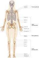
Pectoral and Pelvic Girdles
Pectoral Girdle: Clavicle and scapula, attaches upper extremities.
Upper Extremity: Humerus, ulna, radius, carpals, metacarpals, phalanges.

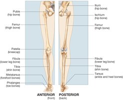
Pelvic Girdle: Ilium, ischium, pubis, attaches lower extremities.
Lower Extremity: Femur, patella, tibia, fibula, tarsals, metatarsals, phalanges.
Joints
Types of Joints
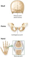
Joints, or articulations, are classified by the movement they allow:
Synovial Joints: Freely moving, most common, e.g., ball-and-socket joints. Contain synovial membrane, ligaments, and sometimes bursae and menisci.
Cartilaginous Joints: Allow slight movement, held by cartilage, e.g., pubic symphysis.
Fibrous Joints: Allow almost no movement, joined by fibrous tissue, e.g., skull sutures.
Structure of a Synovial Joint
Synovial joints have a complex structure:
Joint Capsule: Encloses the joint.
Synovial Membrane: Secretes lubricating fluid.
Articular Cartilage: Covers bone surfaces.
Joint Cavity: Contains synovial fluid.
Bone Pathology and Fractures
Types of Bone Fractures
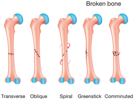
Fractures are classified by their pattern and severity:
Transverse: Straight across the bone.
Oblique: At an angle.
Spiral: Curves around the bone.
Greenstick: Incomplete break, common in children.
Comminuted: Bone is shattered into pieces.
Bone Diseases
Osteogenic Sarcoma: Malignant bone tumor, often seen in the long bones.
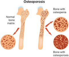
Osteopenia and Osteoporosis: Conditions of reduced bone density, increasing fracture risk.
Spinal Column Pathology
Common Spinal Disorders
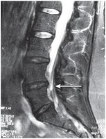
Herniated Disk: Protrusion of intervertebral disk, compressing spinal cord.
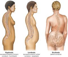
Abnormal Curvatures:
Kyphosis: Excessive posterior thoracic curvature.
Lordosis: Excessive anterior lumbar curvature.
Scoliosis: Lateral curvature of the spine.
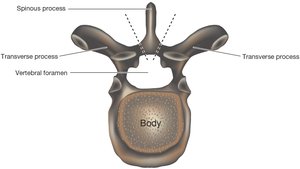
Vertebral Anatomy
Spinous Process: Posterior projection for muscle attachment.
Transverse Process: Lateral projections.
Vertebral Foramen: Opening for spinal cord.
Joint Pathology
Dislocation and Arthritis
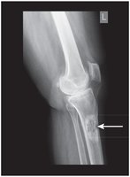
Dislocation: Complete displacement of a bone from its joint.
Osteoarthritis: Degenerative joint disease, bones in direct contact.
Medical Terminology: Key Terms and Word Building
Combining Forms, Suffixes, and Prefixes
Medical terms are constructed from combining forms, suffixes, and prefixes.
Example: Ostealgia = oste/o (bone) + -algia (pain) = bone pain
Example: Synovitis = synov/o (synovial membrane) + -itis (inflammation) = inflammation of a synovial membrane
Pathology Terms
Osteoarthritis: oste/o (bone) + arthr/o (joint) + -itis (inflammation)
Kyphosis: kyph/o (hump) + -osis (abnormal condition)
Summary Table: Types of Joints
Type of Joint | Movement Allowed | Example |
|---|---|---|
Synovial | Freely moving | Shoulder, knee |
Cartilaginous | Slight movement | Pubic symphysis |
Fibrous | Almost no movement | Skull sutures |
Summary Table: Bone Fracture Types
Fracture Type | Description |
|---|---|
Transverse | Straight across the bone |
Oblique | At an angle |
Spiral | Curves around the bone |
Greenstick | Incomplete break |
Comminuted | Bone shattered into pieces |
Key Equations and Terms
Ossification Equation:
Bone Remodeling:
Conclusion
Understanding the musculoskeletal system and its terminology is essential for medical students. Mastery of combining forms, suffixes, and prefixes, as well as anatomical and pathological terms, provides a foundation for clinical practice and further study in medical sciences.
