 Back
BackRespiratory System: Medical Terminology and Anatomy Study Guide
Study Guide - Smart Notes
 Tailored notes based on your materials, expanded with key definitions, examples, and context.
Respiratory System Overview
Introduction and Objectives
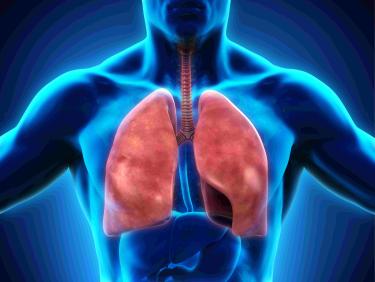
The respiratory system is essential for gas exchange, supplying oxygen to the body and removing carbon dioxide. Understanding its anatomy and physiology is crucial for medical terminology students, as it also interacts closely with the cardiovascular system.
Objective 1: Identify anatomical features of the respiratory system.
Objective 2: Understand basic physiology of respiration.
Objective 3: Comprehend the integration of respiratory and cardiovascular systems.
Functions of the Respiratory System
The respiratory system performs several vital functions to maintain homeostasis.
Inhalation: Intake of fresh air into the lungs.
Gas Exchange: Oxygen is exchanged for carbon dioxide in the lungs.
Exhalation: Removal of stale air and carbon dioxide from the body.
Integration: Works with the cardiovascular system to transport gases.
Respiratory Physiology
Respiration and Its Subdivisions
Respiration is a continuous process required for cellular function. It is divided into distinct phases:
Ventilation: Movement of air between the environment and lungs.
Inhalation: Air flows into the lungs, bringing oxygen to alveoli.
Exhalation: Air flows out, removing carbon dioxide.
External Respiration
External respiration occurs in the lungs, where oxygen and carbon dioxide are exchanged between alveoli and blood.
Oxygen: Diffuses from alveoli into the bloodstream.
Carbon Dioxide: Diffuses from blood into alveoli for exhalation.
Internal Respiration
Internal respiration is the exchange of gases at the cellular level.
Oxygen: Leaves bloodstream, enters tissues, and is used for metabolism.
Carbon Dioxide: Produced by metabolism, enters bloodstream for removal.
Anatomy of the Respiratory System
Major Organs
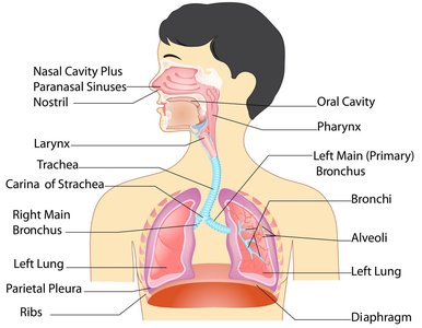
The respiratory system consists of several organs that facilitate air flow and gas exchange:
Nasal cavity
Pharynx
Larynx
Trachea
Bronchial tubes
Bronchi
Lungs
Nasal Cavity
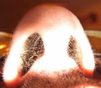
The nasal cavity is the entry point for air and plays a role in filtering, warming, and humidifying inhaled air.
Nares: Air enters through nostrils.
Nasal septum: Divides the cavity.
Palate: Separates nasal cavity from mouth.
Vibrissae: Small hairs filter large particles.
Mucous membrane: Warms and humidifies air.
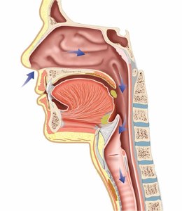
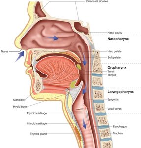
Pharynx
The pharynx, or throat, is a shared passage for air and food. It is divided into three regions:
Nasopharynx: Upper section by nasal cavity.
Oropharynx: Middle section by oral cavity.
Laryngopharynx: Lower section by larynx.
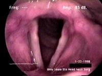
Larynx
The larynx, or voice box, is a muscular tube between the pharynx and trachea. It contains the vocal cords, which vibrate to produce sound.
Cartilage plates: Form the structure, including the Adam's apple (thyroid cartilage).
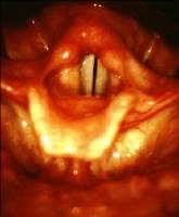
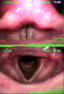
Vocal cords: Folds of tissue that vibrate to produce sound; the opening is called the glottis.
Epiglottis
The epiglottis is a flap of cartilage that covers the larynx and trachea during swallowing, preventing food from entering the airway.
Function: Directs food to the esophagus, not the trachea.
Trachea
The trachea is a tube that carries air from the larynx to the bronchi. It is reinforced by cartilaginous rings and lined with mucous membrane and cilia.
Length: Approximately four inches.
Structure: Smooth muscle, cartilage rings, mucous membrane.
Function: Cleanses, warms, and moistens air.
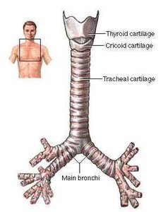
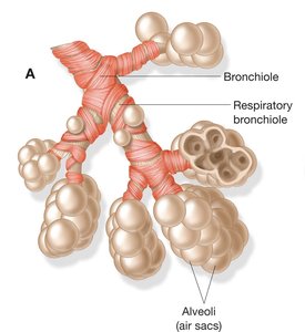
Bronchial Tubes and Bronchi
The distal end of the trachea divides into left and right main bronchi, which branch into secondary and tertiary bronchi, and eventually into bronchioles.
Bronchi: Main passageways into the lungs.
Bronchioles: Narrow branches ending in alveoli.
Alveoli: Air sacs where gas exchange occurs; each lung contains about 150 million alveoli.
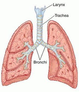
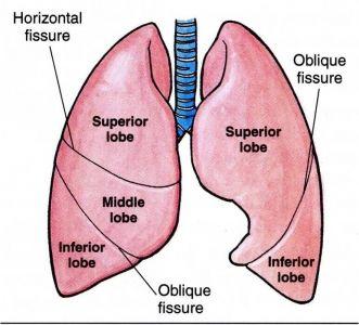
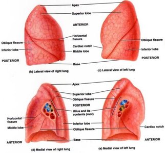
Lungs
The lungs are the primary organs of respiration, consisting of bronchi, bronchioles, and alveoli. The right lung has three lobes, while the left lung has two.
Apex: Pointed superior portion.
Base: Broad lower area.
Hilum: Entry and exit point for bronchi, blood vessels, and nerves.
Protection: Externally protected by the ribs.
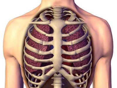
Pleura
The pleura are membranes surrounding the lungs. The parietal pleura lines the chest cavity, while the visceral pleura adheres to the lung surface. Serous fluid between layers reduces friction during ventilation.
Parietal pleura: Outer membrane.
Visceral pleura: Inner membrane.
Serous fluid: Lubricates pleural surfaces.
Respiratory Physiology: Mechanics and Measurements
Pulmonary Function Tests
Pulmonary function tests measure the volume of air flowing in and out of the lungs, helping to determine lung capacity and diagnose respiratory conditions.
Respiratory therapist: Conducts tests and monitors lung function.
Lung Capacities
Lung capacities refer to the different volumes of air the lungs can hold, measured during pulmonary function tests.
Respiratory Muscles
Respiratory muscles, including the diaphragm and intercostal muscles, facilitate inhalation and exhalation.
Inhalation: Diaphragm contracts and moves downward, decreasing pressure in the chest cavity and allowing air to enter.
Exhalation: Diaphragm and intercostal muscles relax, increasing pressure and forcing air out.
Respiratory Rates by Age Group
Respiratory rates vary by age:
Age | Respirations per Minute |
|---|---|
Newborn | 30-60 |
1-year-old | 18-30 |
16-year-old | 16-20 |
Adult | 12-20 |
Medical Terminology and Specialties
Anatomical Terms
Key anatomical terms related to the respiratory system:
Alveolar: Pertaining to the alveoli
Bronchial: Pertaining to a bronchus
Bronchiolar: Pertaining to a bronchiole
Diaphragmatic: Pertaining to the diaphragm
Epiglottic: Pertaining to the epiglottis
Laryngeal: Pertaining to the larynx
Nasal: Pertaining to the nose
Pharyngeal: Pertaining to the pharynx
Pleural: Pertaining to the pleura
Pulmonary: Pertaining to a lung
Septal: Pertaining to the nasal septum
Thoracic: Pertaining to the chest
Tracheal: Pertaining to the trachea
Medical Specialties
Pulmonology: Diagnosis and treatment of respiratory diseases; physician is a pulmonologist.
Thoracic surgery: Surgical treatment of respiratory conditions; physician is a thoracic surgeon.
Respiratory therapy: Allied health specialty assisting patients with respiratory and cardiopulmonary disorders.
Pathology of the Respiratory System
Upper Respiratory System Pathology
Croup: Acute respiratory condition in children; barking cough.
Diphtheria: Bacterial infection; thick membrane across throat.
Laryngitis: Inflammation of the larynx.
Nasopharyngitis: Inflammation of nose and pharynx; common cold.
Pertussis: Bacterial infection; whooping cough.
Bronchial Tube Pathology
Asthma: Difficulty breathing due to bronchospasms, dyspnea, coughing, and wheezing.
Bronchiectasis: Enlarged bronchi from destruction of bronchial wall.
Bronchitis: Inflammation of a bronchus.
Bronchogenic carcinoma: Cancerous tumor originating in bronchi.
Lung Pathology
ARDS: Acute respiratory distress syndrome; respiratory failure.
Anthracosis: Coal dust in lungs; black lung.
Asbestosis: Asbestos fibers in lungs.
Atelectasis: Collapse of alveoli; prevents gas exchange.
COPD: Chronic obstructive pulmonary disease; progressive, irreversible lung conditions.
Cystic fibrosis: Genetic condition; thick mucus causes congestion.
Emphysema: Destruction of alveolar walls.
Histoplasmosis: Fungal infection of lungs.
IRDS: Infant respiratory distress syndrome; common in premature infants.
Influenza: Viral infection of respiratory system.
Pneumocystis pneumonia: Fungal pneumonia; seen in AIDS patients.
Pneumonia: Inflammation; alveoli fill with fluid.
Pulmonary edema: Excess fluid in lung tissues.
Pulmonary embolism: Blood clot obstructs pulmonary artery.
Pulmonary fibrosis: Formation of scar tissue; reduced lung expansion.
SARS: Severe acute respiratory syndrome; high mortality.
Silicosis: Silica dust accumulation in lungs.
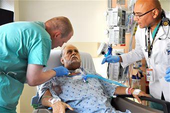
Respiratory Therapy and Procedures
Respiratory Therapy
Nasal cannula: Device to deliver oxygen into the nose.
Postural drainage: Positioning to promote drainage of bronchial secretions.
Supplemental oxygen therapy: Providing additional oxygen concentration.
Ventilator: Machine for artificial ventilation.
Surgical Procedures
Thoracentesis: Surgical puncture to remove fluids from chest wall.
Thoracostomy: Insertion of chest tube to drain fluid or air.
Thoracotomy: Cutting into the chest cavity.
Tracheotomy: Emergency opening in trachea for breathing.
Respiratory System Pharmacology
Medications
Bronchodilator: Relaxes bronchospasms; treats asthma (e.g., Proventil, Ventolin, Theo-Dur).
Corticosteroids: Reduces inflammation (e.g., Flonase, Nasonex, Azmacort).
Decongestant: Reduces congestion (e.g., Afrin, Drixoral, Sudafed).
Expectorant: Improves ability to cough up mucus (e.g., Robitussin, Mucinex).
Mucolytic: Liquefies mucus (e.g., Mucomyst).
Abbreviations in Respiratory System
Abbreviation | Meaning |
|---|---|
ABGs | Arterial blood gases |
ARDS | Adult respiratory distress syndrome |
Bronch | Bronchoscopy |
CO2 | Carbon dioxide |
COPD | Chronic obstructive pulmonary disease |
CPR | Cardiopulmonary resuscitation |
C&S | Culture and sensitivity |
Key Equations
Gas Exchange Formula
The partial pressure of gases determines their diffusion:
Respiratory Rate Calculation
Minute ventilation is calculated as:
Additional info: Academic context was added to clarify anatomical terms, pathology, and pharmacology for completeness and exam preparation.
