 Back
BackRespiratory System: Medical Terminology and Clinical Concepts
Study Guide - Smart Notes
 Tailored notes based on your materials, expanded with key definitions, examples, and context.
Pulmonology: Medical Terminology
Definition and Word Parts
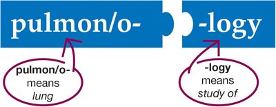
Pulmonology is the medical specialty focused on the study of the respiratory system, including its anatomy, physiology, diseases, and treatments. Understanding medical terminology is essential for proficiency in this area.
pulmon/o-: means lung
-logy: means study of
Respiratory System Overview
Major Structures
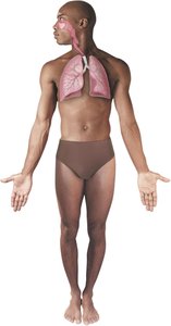
The respiratory system consists of the lungs and associated structures that form a pathway for air to flow into and out of the body. It is divided into upper and lower respiratory tracts.
Lungs: Main organs responsible for gas exchange
Pathways: Nose, pharynx, larynx, trachea, bronchi, bronchioles, alveoli
Anatomy: Upper Respiratory System
Nose and Nasal Cavity
The nose is the entry point for air and contains the nasal cavity, which is divided by the septum. Turbinates slow airflow, allowing air to be warmed and moistened. The nasal mucosa lines the cavity, producing mucus to humidify air and trap particles.
Turbinates: Slow airflow for warming and moistening
Nasal mucosa: Humidifies air, produces mucus
Cilia: Move mucus and debris out of the nasal cavity
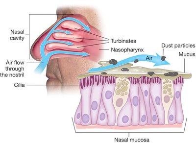
Air Filtration and Humidification
Air entering the nasal cavity swirls around the turbinates, allowing the mucosa to warm and moisten the air. Cilia move in waves to clear away mucus and inhaled debris, helping maintain core body temperature and protect the lungs.
Mucus and hair: Trap particles and prevent them from reaching the lungs
Importance: Especially critical in cold or dry environments

Anatomy: Upper Respiratory System
Pharynx
The pharynx is a common passage for inhaled air, exhaled air, and swallowed food. It has three parts: nasopharynx, oropharynx, and laryngopharynx.
Nasopharynx: Uppermost part, behind the nasal cavity
Oropharynx: Middle part, behind the oral cavity
Laryngopharynx: Lowest part, leading to the larynx and esophagus

Anatomy: Lower Respiratory System
Larynx
The larynx, or voice box, remains open during breathing and contains the vocal cords. During swallowing, the larynx moves up to the epiglottis, preventing food from entering the lungs.
Vocal cords: Produce sound when air is exhaled
Epiglottis: Covers the larynx during swallowing
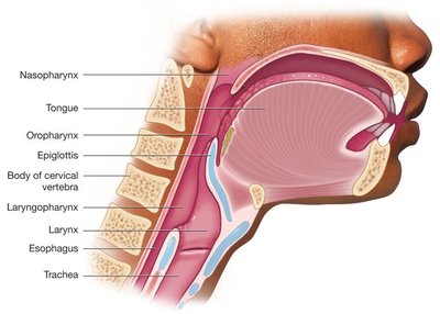
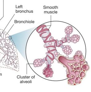
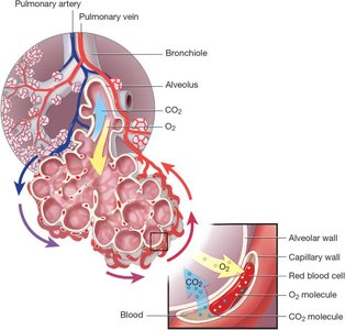
Trachea, Bronchi, Bronchioles, and Alveoli
The trachea (windpipe) is a passageway for air, supported by C-shaped cartilage rings. It divides into right and left bronchi, which enter the lungs and branch into bronchioles. Bronchioles end in alveoli, clusters of microscopic air sacs where gas exchange occurs.
Trachea: Rigid anteriorly, soft posteriorly
Bronchi: Main branches entering the lungs
Bronchioles: Smaller branches within the lungs
Alveoli: Sites of oxygen and carbon dioxide exchange
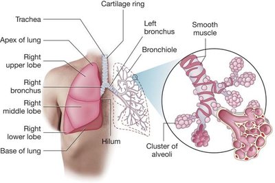
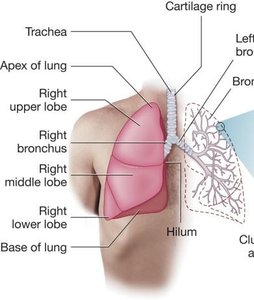
Lungs
The lungs are spongy, air-filled structures. The right lung has three lobes (upper, middle, lower), while the left lung has two lobes (upper, lower). The apex is the rounded top of the lung.
Right lung: RUL, RML, RLL
Left lung: LUL, LLL
Apex: Top of the lung
Alveoli and Pulmonary Parenchyma
Inside the lung, bronchi branch into bronchioles ending in alveoli. Alveoli are hollow spheres of cells that exchange oxygen and carbon dioxide with capillaries. They secrete surfactant to prevent collapse and collectively form the pulmonary parenchyma.
Surfactant: Reduces surface tension, prevents alveolar collapse
Pulmonary parenchyma: Functional tissue of the lung
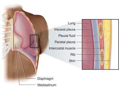
Thorax and Pleura
The thorax is the area between the base of the neck and the diaphragm. The rib cage forms the bony wall, and the mediastinum lies between the lungs. The diaphragm is the inferior border. The pleurae are membranes that cover the lungs and line the thoracic cavity, secreting pleural fluid.
Visceral pleura: Covers the lung surface
Parietal pleura: Lines the thoracic cavity
Pleural fluid: Lubricates pleural surfaces
Physiology: Breathing and Respiration
Breathing Mechanisms
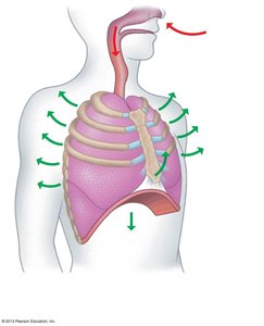
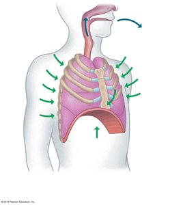
Breathing is involuntary, regulated by respiratory control centers in the brain. The phrenic nerve stimulates the diaphragm. Inhalation (inspiration) and exhalation (expiration) are the two main phases.
Inhalation: Diaphragm contracts, thoracic cavity enlarges, air flows in
Exhalation: Diaphragm relaxes, thoracic cavity shrinks, air flows out
Processes of Respiration
Respiration involves five processes:
Ventilation: Movement of air in and out of the lungs
External respiration: Exchange of gases between blood and lungs
Gas transport: Movement of gases by the blood
Internal respiration: Movement of oxygen from blood to cells
Cellular respiration: Use of oxygen by cells to generate energy; production of carbon dioxide as waste
Diseases of the Respiratory System
Upper Respiratory Diseases
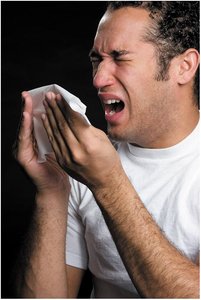
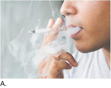
Upper respiratory infection (URI) is a bacterial or viral infection of the upper respiratory tract, commonly known as a head cold. It spreads by contact or inhalation of particles.
Antibiotics: Effective against bacterial infections
Transmission: By droplets or contact
Lower Respiratory Diseases
Common diseases include asthma, bronchiectasis, bronchitis, and chronic obstructive pulmonary disease (COPD). Asthma is characterized by hyperreactivity of bronchi, inflammation, excess mucus, and bronchospasm. COPD includes chronic bronchitis and emphysema, often due to smoking or pollution.
Asthma: Bronchospasm, inflammation, excess mucus
Bronchiectasis: Permanent enlargement of bronchioles
Bronchitis: Infection or inflammation of bronchi
COPD: Chronic inflammation, severe coughing, shortness of breath
Lung Diseases
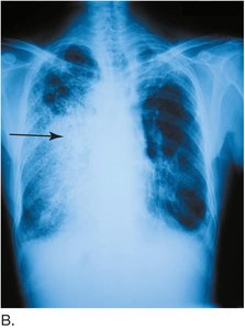
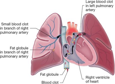
Diseases affecting the lungs include pneumonia, pulmonary edema, pulmonary embolism, tuberculosis, and cystic fibrosis. Pneumonia is an infection of the lung lobes, causing fluid and white blood cells to fill passages. Pulmonary embolism is a blockage of a pulmonary artery by an embolus.
Pneumonia: Bacterial, viral, aspiration, lobar, panlobar, pneumococcal, walking pneumonia
Pulmonary edema: Fluid buildup in alveoli
Pulmonary embolism: Blockage by blood clot or fat globule
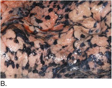
Tuberculosis: Caused by Mycobacterium tuberculosis, forms nodules in lungs
Cystic fibrosis: Inherited, thick mucus blocks alveoli, frequent infections
Laboratory Tests and Diagnostic Procedures
Common Tests
Several laboratory and diagnostic tests are used to assess respiratory function and diagnose diseases.
Arterial blood gases (ABGs): Measures oxygen and carbon dioxide in arterial blood
Oximetry: Measures oxygen saturation of hemoglobin
Pulmonary function test (PFT): Measures lung volumes and capacities
Sputum culture and sensitivity (C&S): Identifies causative bacteria and antibiotic sensitivity
Tuberculosis tests: Tine, Mantoux, acid-fast bacillus, Xpert sputum
Radiologic and Nuclear Medicine Procedures
Imaging Techniques
Imaging is essential for diagnosing respiratory diseases.
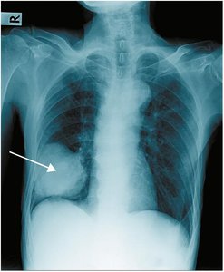
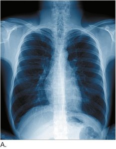
Chest radiography (CXR): X-ray imaging of the lungs
Computerized axial tomography (CAT, CT): Cross-sectional imaging
Lung scan: Identifies areas of poor gas uptake
Magnetic resonance imaging (MRI): Uses magnetic fields and radio waves
Medical and Surgical Procedures
Common Procedures
Medical and surgical procedures are used to treat respiratory diseases and manage airway function.
Auscultation and percussion (A&P): Listening to breath sounds and tapping the back
Cardiopulmonary resuscitation (CPR): Emergency procedure for cardiac and respiratory arrest
Chest percussion therapy: Loosens mucus in the lungs
Continuous positive airway pressure (CPAP): Treats sleep apnea
Endotracheal intubation: Establishes airway with a tube
Incentive spirometry: Encourages deep breathing post-surgery
Nebulizer treatment: Delivers medication as a mist
Oxygen therapy: Provides supplemental oxygen
Bronchoscopy: Examines trachea and bronchi
Chest tube insertion: Removes air, fluid, pus, or blood
Lung resection: Removes part or all of a lung
Thoracentesis: Removes pleural fluid
Thoracotomy: Incision into thoracic cavity
Tracheostomy: Provides access to lungs for respiratory support
Abbreviation Summary
Common Respiratory Abbreviations
Abbreviation | Definition |
|---|---|
A&P | auscultation and percussion |
ABG | arterial blood gases |
CXR | chest x-ray |
COPD | chronic obstructive pulmonary disease |
CPAP | continuous positive airway pressure |
CF | cystic fibrosis |
MDI | metered-dose inhaler |
PFT | pulmonary function test |
TB | tuberculosis |
URI | upper respiratory infection |
Key Medical Terms and Definitions
Bronchopulmonary: Refers to bronchi, bronchioles, and lungs
Dyspnea: Difficult or labored breathing
Apnea: Absence of breathing
Tachypnea: Abnormally fast breathing
Cyanosis: Bluish skin due to low oxygen
Hemothorax: Blood in the thoracic cavity
Pneumothorax: Air in the pleural space causing lung collapse
Summary Table: Respiratory Diseases
Disease | Definition | Key Features |
|---|---|---|
Asthma | Hyperreactivity of bronchi | Inflammation, bronchospasm, excess mucus |
COPD | Chronic bronchitis/emphysema | Chronic inflammation, barrel chest |
Pneumonia | Infection of lung lobes | Fluid, WBCs, microorganisms in passages |
Pulmonary embolism | Blockage of pulmonary artery | Blood clot, reduced oxygenation |
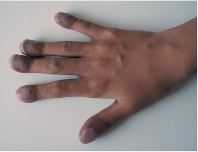
Cystic fibrosis | Inherited thick mucus disorder | Frequent infections, clubbing of fingers |
Tuberculosis | Bacterial lung infection | Nodules, resistant to single drug |
