 Back
BackSkeletal System: Structure, Function, and Pathology
Study Guide - Smart Notes
 Tailored notes based on your materials, expanded with key definitions, examples, and context.
Skeletal System Overview
Functions of the Skeletal System
The human skeleton is a complex framework consisting of 206 bones that provides structure, protection, and support for the body. It plays a vital role in movement, mineral storage, and blood cell formation.
Support: The skeleton forms the supporting framework of the body.
Protection: Bones protect vital internal organs (e.g., skull protects the brain, rib cage protects the heart and lungs).
Movement: Bones serve as attachment points for muscles, enabling movement.
Mineral Storage: Bones act as reservoirs for minerals such as calcium and phosphorus.
Blood Cell Formation: Red bone marrow within certain bones produces blood cells (hematopoiesis).
Classification and Structure of Bones
Bone Classification by Shape
Bones are classified according to their shapes, which relate to their functions:
Long bones: e.g., femur, humerus
Short bones: e.g., carpals, tarsals
Flat bones: e.g., sternum, scapula
Irregular bones: e.g., vertebrae
Sesamoid bones: e.g., patella
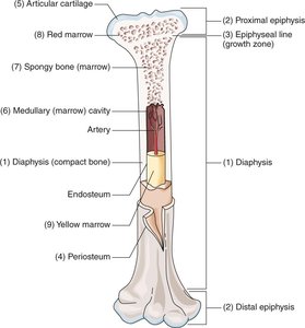
Structure of a Long Bone
Long bones have specialized regions and tissues that contribute to their function and growth.
Diaphysis: The main shaft of the bone, composed of compact bone for strength.
Epiphysis: The ends of the bone, which articulate with other bones.
Epiphyseal Line: Cartilaginous layer that allows for lengthwise bone growth during development.
Periosteum: Fibrous membrane covering the bone's surface, containing nerves and blood vessels.
Articular Cartilage: Smooth cartilage covering joint surfaces to reduce friction.
Medullary (Marrow) Cavity: Central cavity containing yellow marrow (fat storage in adults).
Spongy (Cancellous) Bone: Porous bone tissue found mainly in the epiphyses, containing red marrow for blood cell production.
Red Bone Marrow: Site of hematopoiesis (blood cell formation).
Yellow Marrow: Stores fat; not active in blood cell formation in adults.
Bone Formation and Growth
Ossification and Bone Cells
Bone formation, or ossification, begins before birth and continues through adolescence. It involves the replacement of cartilage and connective tissue with bone.
Osteoblasts: Cells that actively produce new bone tissue.
Osteoclasts: Cells that digest and break down bone tissue.
Osteocytes: Mature bone cells that maintain bone tissue.
Ossification is essential for bone growth, repair, and remodeling throughout life.
Bone Markings and Depressions
Types of Bone Markings
Bones have various markings that serve as attachment points, passageways, and articulations:
Enlargements: Projections for muscle and tendon attachment.
Openings: For passage of nerves and blood vessels (e.g., foramen).
Depressions: Concave areas that help form joints or serve as muscle attachment points.
Common Bone Depressions
Sulcus: Groove or depression (e.g., fissure).
Sinus: Hollow space within a bone (e.g., frontal sinus).
Fossa: Shallow depression (e.g., iliac fossa).
Foramen: Hole for passage of nerves or blood vessels (e.g., foramen magnum of the skull).
Axial Skeleton: Skull and Vertebral Column
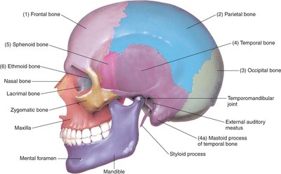
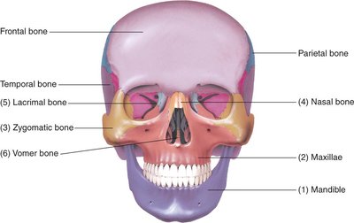
Cranial and Facial Bones
The skull protects the brain and forms the structure of the face. It consists of cranial and facial bones joined by immovable joints called sutures.
Cranial Bones: Eight bones forming the cranium (e.g., frontal, parietal, temporal, occipital, sphenoid, ethmoid).
Facial Bones: Fourteen bones, including the maxillae (upper jaw) and mandible (lower jaw). The mandible is the only movable facial bone.
Vertebral Column
The vertebral column, or spine, forms the body's central axis and protects the spinal cord. It consists of 24 vertebrae, the sacrum, and the coccyx.
Cervical Vertebrae (C1–C7): Neck region; C1 (atlas) and C2 (axis) are specialized for head movement.
Thoracic Vertebrae (T1–T12): Chest region; articulate with ribs.
Lumbar Vertebrae (L1–L5): Lower back; largest and strongest vertebrae.
Sacrum: Triangular bone formed by fusion of five sacral vertebrae.
Coccyx: Tailbone; formed by fusion of four coccygeal bones.
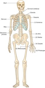
Thorax and Appendicular Skeleton
Bones of the Thorax
Ribs: Twelve pairs classified as true (1–7), false (8–10), and floating (11–12) based on their anterior attachments.
Sternum: Breastbone, consisting of the manubrium, body, and xiphoid process.
Clavicle: Collarbone, connecting the sternum to the scapula.
Scapula: Shoulder blade, articulates with the clavicle and humerus.
Bones of the Upper Extremities
Humerus: Upper arm bone.
Radius: Forearm bone on the thumb side.
Ulna: Forearm bone on the little finger side.
Carpals: Wrist bones.
Metacarpals: Hand bones.
Phalanges: Finger bones (three per finger, two in the thumb).
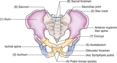
Pelvic Bones
Ilium: Largest, uppermost hip bone.
Iliac Crest: Upper curved edge of the ilium.
Ischium: Lowest, strongest part of the hip bone.
Pubis: Anterior part of the hip bone.
Symphysis Pubis: Cartilaginous joint connecting the two pubic bones.
Acetabulum: Socket for the femur, formed by the ilium, ischium, and pubis.
Sacrum and Coccyx: Posterior pelvic bones.
Bones of the Lower Extremities
Femur: Thigh bone; longest and strongest bone in the body.
Patella: Kneecap.
Tibia: Shin bone; larger and stronger lower leg bone.
Fibula: Slender lower leg bone.
Tarsals: Ankle bones (including calcaneus and talus).
Metatarsals: Foot bones.
Phalanges: Toe bones (three per toe, two in the great toe).
Pathological Conditions of the Skeletal System
Common Disorders
Osteomalacia: Softening of bones due to calcium and phosphorus deficiency; called rickets in children.
Osteomyelitis: Infection of bone and bone marrow, usually bacterial.
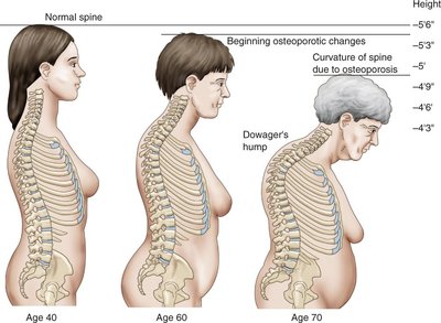
Osteoporosis: Loss of bone density, making bones fragile and prone to fractures.
Osteochondroma: Most common benign bone tumor, often affecting femur and tibia.
Paget’s Disease (Osteitis Deformans): Nonmetabolic bone disease with excessive, disorganized bone formation.
Spinal Stenosis: Narrowing of the vertebral canal, causing nerve compression.
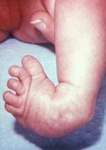
Talipes Equinovarus (Clubfoot): Congenital deformity with foot fixed in plantar flexion and medial deviation.
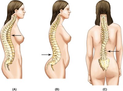
Abnormal Curvatures of the Spine
Kyphosis: Outward (posterior) curvature, resulting in a hunchback appearance.
Lordosis: Inward (anterior) curvature, resulting in swayback.
Scoliosis: Lateral (sideward) curvature of the spine.
Fractures: Types and Treatment
Types of Fractures
Closed (Simple) Fracture: Bone is broken but skin is intact.
Open (Compound) Fracture: Bone breaks through the skin.
Complete Fracture: Break extends through the entire bone.
Incomplete (Greenstick) Fracture: Bone is partially broken and bent; common in children.
Compression Fracture: Bone surfaces are forced against each other.
Impacted Fracture: Broken ends of bone are forced into each other.
Comminuted Fracture: Bone is splintered or crushed.
Colles’ Fracture: Break at the distal radius near the wrist.
Hairline (Stress) Fracture: Minor fracture with bone alignment maintained.
Pathological Fracture: Occurs in bone weakened by disease.
Treatment of Fractures
Closed Reduction: Non-surgical realignment of bone fragments.
Open Reduction: Surgical intervention to align and fix bone fragments.
Diagnostic Techniques, Treatments, and Procedures
Diagnostic Techniques
Bone Scan: Intravenous radioisotope injection followed by skeletal imaging to detect abnormal bone metabolism.
Bone Marrow Aspiration: Needle removal of bone marrow for microscopic examination; used to diagnose blood disorders.
Bone Density Testing:
Dual Energy X-ray Absorptiometry (DEXA): Most common, noninvasive test for osteopenia and osteoporosis.
Dual Photon Absorptiometry and Quantitative Computed Tomography (QCT): Less commonly used methods.
Example: DEXA scan is preferred for osteoporosis screening due to its accuracy, speed, and low radiation exposure.
