 Back
BackSkeletal System: Structure, Function, Diseases, and Medical Terminology
Study Guide - Smart Notes
 Tailored notes based on your materials, expanded with key definitions, examples, and context.
Orthopedics and Medical Terminology
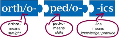
Definition and Etymology
Orthopedics is the medical specialty focused on the anatomy and physiology of the skeletal and muscular systems. The term is derived from Greek roots:
orth/o-: means straight
ped/o-: means child
-ics: means knowledge; practice
Orthopedists use diagnostic tests, medical and surgical procedures, and drugs to treat skeletal and muscular diseases.
Skeletal System Overview
Structure and Organization
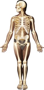
The skeletal system is a widespread, connected body system consisting of 206 bones and other structures. It extends from the top of the head to the tips of the fingers and toes.
Axial skeleton: Central structure including the head, neck, chest, and spinal bones.
Appendicular skeleton: Limbs, including the shoulder, arm, hip, and leg bones.
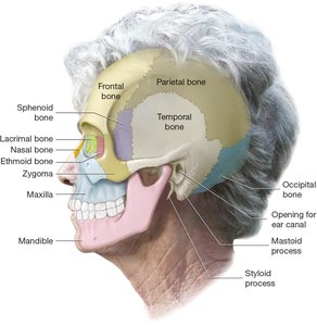
Bones of the Skull
Lateral View
The skull consists of cranial and facial bones. The lateral view shows:
Frontal bone: Forms the forehead
Parietal bones: Form the upper sides and posterior
Occipital bone: Forms the base
Temporal bones: Form the sides
Sphenoid bone: Forms the central base
Ethmoid bone: Forms the nasal septum
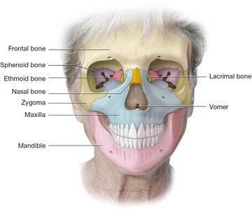
Frontal View
The facial bones support the nose, cheeks, and lips. The mandible is the only moveable bone of the skull. The palatine bones fuse to form the hard palate (not shown).
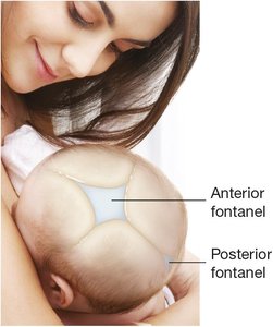
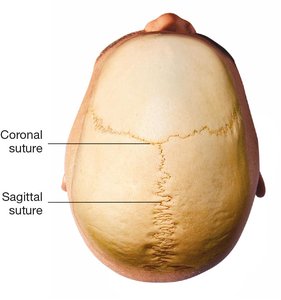
Fontanels
Fontanels are soft spots on an infant's skull where the bones have not yet fused. The anterior fontanel is between the two frontal bones, and the posterior fontanel is between the two parietal bones. These areas are covered by fibrous connective tissue.
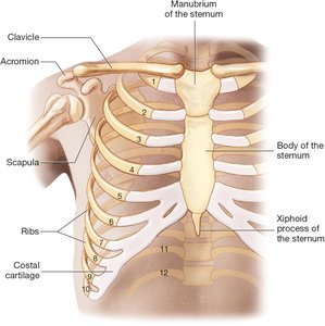
Bones of the Chest and Shoulder
Thorax and Shoulder Girdle
The sternum and ribs form the thorax, a bony cage that protects the heart and lungs. The clavicle and scapula are bones of the shoulder.
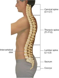
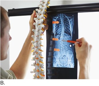
Bones of the Spine
Vertebral Column
The spine supports the head, neck, and trunk and protects the spinal cord. It consists of five regions:
Cervical vertebrae (C1–C7)
Thoracic vertebrae (T1–T12)
Lumbar vertebrae (L1–L5)
Sacrum
Coccyx
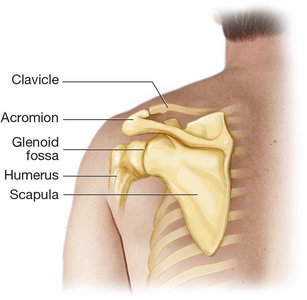
Bones of the Upper Extremity
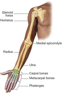
Shoulder and Arm
The scapula joins the humerus at the glenoid fossa. The acromion of the scapula connects to the clavicle, allowing free movement of the shoulder.
Forearm, Wrist, and Hand
The humerus joins with the radius and ulna. The radius and ulna rotate around each other, allowing the hand to turn. The wrist contains eight carpal bones, the hand has five metacarpal bones, and each finger has three phalanges (except the thumb, which has two).
Bones of the Hip and Lower Extremity
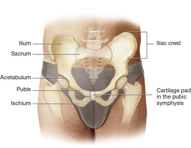
Hip Bones
The ilium and ischium on each side of the hip flow into each other, with the pubis containing a symphysis joint.
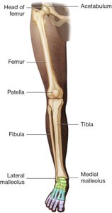
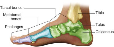
Leg, Ankle, and Foot
The femur joins the tibia to support body weight. The fibula is the smaller bone of the lower leg. The patella protects the knee joint. The tarsal bones in the ankle connect to the metatarsal bones in the foot, and each toe contains three phalanges (except the great toe, which has two).
Joints, Cartilage, and Ligaments
Types of Joints
Joints (articulations) are formed where two bones meet. Types include:
Suture joints: Immovable, found between cranial and facial bones.
Symphysis joints: Slightly moveable, found at the symphysis pubis and vertebrae.
Synovial joints: Fully moveable, including hinge and ball-and-socket joints.
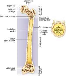
Structure of a Bone
Osseous tissue is a type of connective tissue. The surface of a bone is covered with periosteum. A long bone has a diaphysis (shaft) and epiphyses (ends). The internal structure includes dense cortical bone, a medullary cavity with yellow marrow, and cancellous bone filled with red marrow.
Bone and Cartilage Diseases
Types of Fractures
Fractures are classified by the pattern and cause of the break:
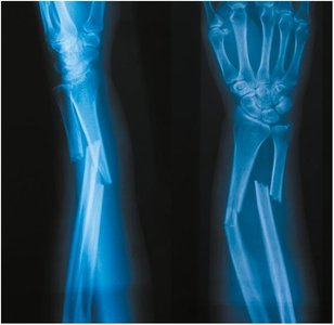
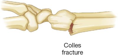
Colles fracture: Distal radius broken by falling on an outstretched hand.
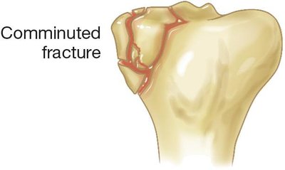
Comminuted fracture: Bone is crushed into several pieces.
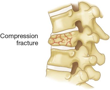
Compression fracture: Vertebrae are compressed together.
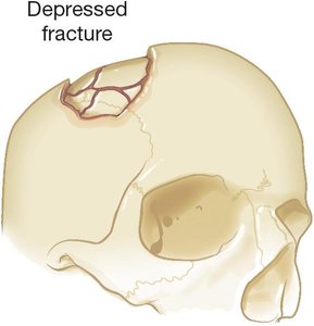
Depressed fracture: Cranium is fractured inward.
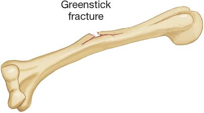
Greenstick fracture: Bone is broken on one side, common in children.
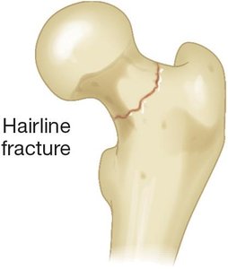
Hairline fracture: Very thin fracture line, bone pieces still together.
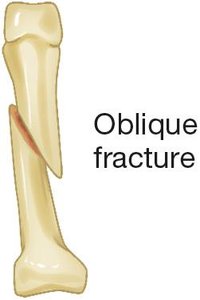
Oblique fracture: Bone is broken at an oblique angle.
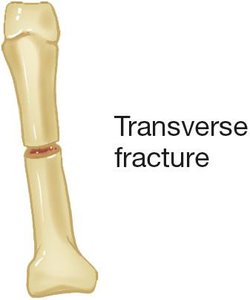
Transverse fracture: Bone is broken perpendicular to its long axis.
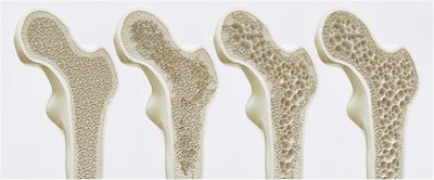
Osteoporosis
Osteoporosis is abnormal thinning of bone structure due to bone breakdown exceeding new bone formation, resulting in demineralization and increased fracture risk.
Spinal and Lower Extremity Deformities
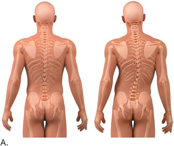
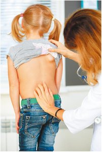
Scoliosis
Scoliosis is an abnormal, excessive, C-shaped or S-shaped lateral curvature of the spine. It can impair movement, posture, and breathing.
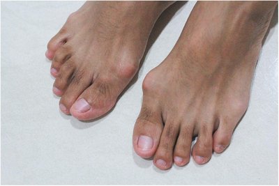
Hallux Valgus and Clubfoot
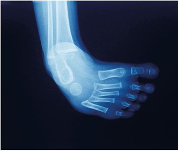
Hallux valgus is a deformity where the great toe is angled laterally, often resulting in a bunion. Clubfoot (talipes equinovarus) is a congenital deformity where the foot is pulled downward and inward.
Joint Diseases
Osteoarthritis and Rheumatoid Arthritis
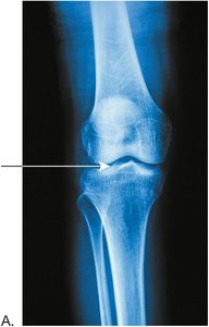
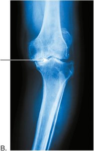
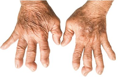
Osteoarthritis is a chronic inflammatory disease of joints, especially weight-bearing joints, characterized by cartilage degeneration and bone spur formation. Rheumatoid arthritis is an autoimmune disorder causing inflammation and deformity of joints, most often in the hands and feet.
Diagnostic and Radiologic Procedures
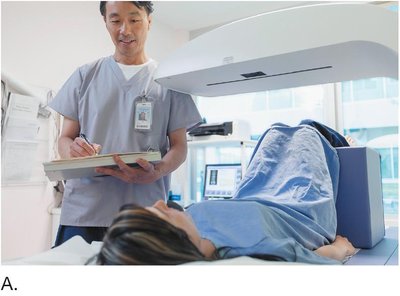
Bone Densitometry
Bone density tests measure bone mineral density to assess for osteoporosis. The hip and spine are optimal sites for testing.
Key Abbreviations
Abbreviation | Definition |
|---|---|
AKA | Above-the-knee amputation |
BKA | Below-the-knee amputation |
BMD | Bone mineral density |
DJD | Degenerative joint disease |
NSAID | Nonsteroidal anti-inflammatory drug |
ORIF | Open reduction and internal fixation |
RA | Rheumatoid arthritis |
ROM | Range of motion |
THR | Total hip replacement |
