 Back
BackTechnical Aspects of the EKG: Medical Terminology and Clinical Application
Study Guide - Smart Notes
 Tailored notes based on your materials, expanded with key definitions, examples, and context.
Technical Aspects of the EKG
Introduction to EKG Recording
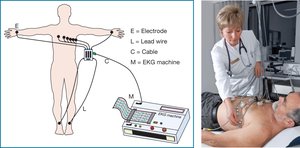
The electrocardiogram (EKG or ECG) is a diagnostic tool that records the electrical activity of the heart. The heart acts as a transmitter of electrical impulses, which are detected by electrodes placed on the body. These impulses are transmitted through lead wires and cables to the EKG machine, where they are amplified and converted from analog to digital signals for interpretation.
Electrodes: Sensors placed on the skin to detect electrical activity.
Lead wires and cables: Conduct the electrical signal from the electrodes to the EKG machine.
Amplifier: Increases the strength of the electrical signal.
Digital converter: Changes the analog signal into a digital format for display and analysis.
Control Features of the EKG Machine
EKG machines have several control features that affect the quality and interpretation of the tracing:
Chart speed: Regulates the speed of the EKG printout. The standard speed is 25 mm/second.
Gain: Adjusts the amplitude (height) of the EKG waves and complexes. The default setting is 10 mm/mV. Increasing the gain may be necessary if the tracing appears flat.
Frequency response: Filters out extraneous noise and artifact to improve tracing clarity.
Electrical Safety in EKG
Proper electrical safety is essential to prevent harm to patients during EKG procedures. Two types of electrical shock are possible:
Macroshock: A high-voltage shock (e.g., 110 volts) due to inadequate grounding, frayed wires, or damaged outlets. Can cause burns, neurologic damage, or fatal arrhythmias.
Microshock: A small shock that can travel through a conduit (such as a pacemaker lead) directly to the heart, potentially causing dangerous arrhythmias even at low voltages.
Precaution: Always check for frayed wires or damaged components before performing an EKG.
Artifact in EKG Recordings
Definition and Types of Artifact
Artifact refers to unwanted interference or jitter on the EKG tracing that does not originate from the heart's electrical activity. Recognizing and correcting artifact is crucial for accurate diagnosis. There are four main types:
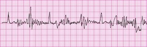
Somatic tremors: Jittery patterns caused by patient movement or shaking wires.
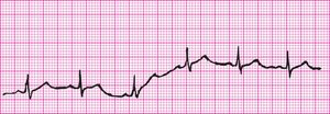
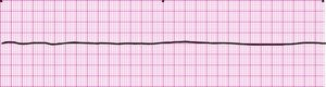
Baseline sway: Up-and-down movement of the baseline, often due to breathing patterns or substances (e.g., lotion, sweat) interfering with electrode contact.
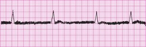
60-cycle interference: Thickened tracing caused by electrical devices nearby. Modern machines have filters to reduce this artifact.
Broken recording: Caused by a frayed or fractured wire, or a loose electrode or cable, resulting in a disconnected or interrupted tracing.
Troubleshooting Artifact
Troubleshooting involves identifying and correcting the source of artifact or recording errors. Steps include:
Identify the common limb or lead affected and focus corrective efforts there.
Replace electrode patches or reattach loose wires as needed.
If artifact is present on a hospital monitor, switch to a different lead (e.g., V1 or MCL1) to obtain a clearer rhythm.
Artifact Masquerading as Cardiac Rhythms
Artifact Mimicking Life-Threatening Rhythms
Artifact can sometimes resemble dangerous cardiac rhythms, leading to unnecessary emergency responses. It is essential to verify the patient's condition before acting on suspicious tracings.
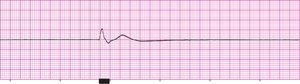
Artifact masquerading as asystole (flat line): Often caused by loose monitor patches or wires.
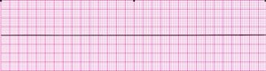
True asystole: Indicates no cardiac electrical activity; the patient will have no pulse and will not be breathing.
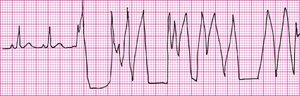
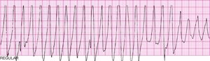
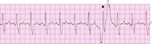
Toothbrush tachycardia: Arm movements during activities like brushing teeth can create artifact that mimics a lethal rhythm.
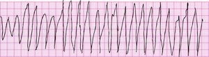
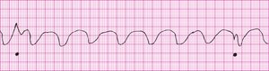
True ventricular tachycardia: A real, life-threatening arrhythmia that must be distinguished from artifact.
CPR artifact: Chest compressions during CPR can produce tracings that resemble ventricular rhythms.
Rhythm without CPR artifact: When CPR is paused, the underlying rhythm can be assessed more accurately.
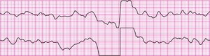
Defibrillation/Cardioversion artifact: Electrical surges from defibrillation or cardioversion cause abrupt changes in the tracing, followed by the return of the true rhythm.
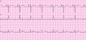
Artifact in multiple leads: Simultaneous recording in several leads can help distinguish artifact (which may affect some leads more than others) from true rhythms.
How to Distinguish Real Rhythms from Artifact
To determine whether a tracing represents a true cardiac rhythm or artifact, follow these steps:
Check the patient for symptoms (e.g., pulse, breathing).
Assess whether the rhythm meets normal criteria for that arrhythmia.
Check another lead, preferably V1 or MCL1, which are less prone to muscle artifact.
Inspect wires and patches for looseness or detachment.
Observe for muscle movements or twitches that could cause artifact.
Follow the QRS complexes, if visible, to help differentiate artifact from true electrical activity.
Summary Table: Types of EKG Artifact
Type of Artifact | Appearance | Common Cause | Correction |
|---|---|---|---|
Somatic tremors | Jittery, irregular baseline | Patient movement, shivering | Calm patient, secure wires |
Baseline sway | Wandering baseline | Breathing, lotion/sweat | Clean skin, proper electrode placement |
60-cycle interference | Thick, fuzzy baseline | Nearby electrical devices | Remove devices, use filters |
Broken recording | Interrupted or flat line | Loose/frayed wires, detached electrodes | Replace/reattach components |
