 Back
BackThe Cardiovascular System: Structure, Function, and Medical Terminology
Study Guide - Smart Notes
 Tailored notes based on your materials, expanded with key definitions, examples, and context.
Cardiovascular System Overview
Introduction to the Cardiovascular System
The cardiovascular system, also known as the circulatory system, is responsible for maintaining the distribution of blood throughout the body. It delivers oxygen and nutrients to cells and removes carbon dioxide and other waste products for elimination by the lungs, liver, and kidneys. The system is composed of the heart and blood vessels, which include arteries, capillaries, and veins. Circulation is divided into two main circuits: systemic circulation and pulmonary circulation.
Systemic circulation: Carries oxygenated blood from the left side of the heart to the body and returns deoxygenated blood to the right side of the heart.
Pulmonary circulation: Carries deoxygenated blood from the right side of the heart to the lungs and returns oxygenated blood to the left side of the heart.
Anatomy of the Heart
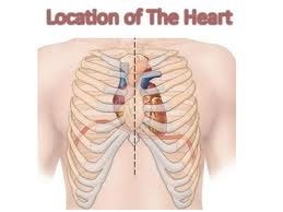
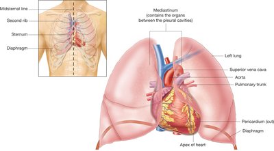
Structure and Location
The heart is a muscular pump made up of cardiac muscle fibers. It is located in the mediastinum, slightly to the left of the chest, behind the sternum. The heart is about the size of a fist and shaped like an upside-down pear, with the tip (apex) pointing downward.
Beats an average of 60–100 times per minute (bpm), or about 100,000 times a day.
Pumps approximately 4,000 gallons of blood per day.
Heart Layers
The heart wall is composed of three layers:
Endocardium: Inner layer lining the heart chambers; smooth and thin to reduce friction.
Myocardium: Thick, muscular middle layer responsible for contraction and generating pressure to pump blood.
Epicardium: Outer layer forming the visceral layer of the pericardial sac; fluid between layers reduces friction as the heart beats.
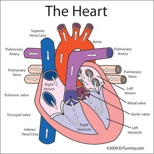
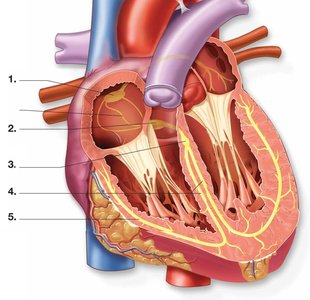
Heart Chambers
The heart is divided into four chambers:
Atria (right and left): Upper receiving chambers; blood returns to the atria via veins (superior/inferior vena cava and pulmonary veins).
Ventricles (right and left): Lower pumping chambers; blood exits the ventricles into arteries (aorta and pulmonary artery).
The heart is divided into right and left sides by the septum.
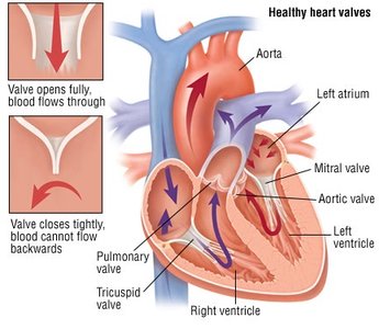
Heart Valves
There are four valves in the heart that act as gates to control the direction of blood flow and prevent backflow:
Tricuspid valve (right atrioventricular valve)
Pulmonary valve (right semilunar valve)
Mitral (bicuspid) valve (left atrioventricular valve)
Aortic valve (left semilunar valve)
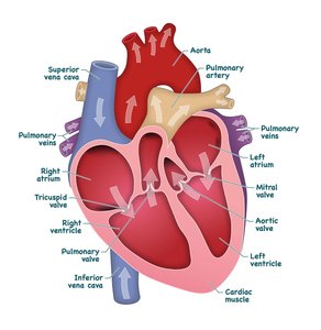
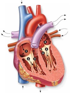
Path of Blood Flow Through the Heart
Stepwise Blood Flow
The path of blood through the heart ensures that deoxygenated blood is sent to the lungs for oxygenation and oxygenated blood is delivered to the body:
Deoxygenated blood from the body enters the right atrium via the superior and inferior vena cava.
Right atrium contracts, pushing blood through the tricuspid valve into the right ventricle.
Right ventricle contracts, sending blood through the pulmonary valve into the pulmonary artery and to the lungs.
Oxygenated blood returns from the lungs to the left atrium via the pulmonary veins.
Left atrium contracts, pushing blood through the mitral valve into the left ventricle.
Left ventricle contracts, sending blood through the aortic valve into the aorta and out to the body.
Cardiac Cycle: Systole and Diastole
Phases of the Cardiac Cycle
The heart alternates between two main phases:
Diastole: Relaxation phase; chambers fill with blood.
Systole: Contraction phase; blood is pushed forward.
Blood pressure is measured as systolic over diastolic pressure (e.g., 120/80 mmHg).
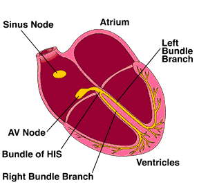
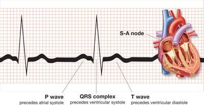
Conduction System of the Heart
Electrical Pathway
The heart's rhythm is controlled by specialized tissue that generates and conducts electrical impulses:
Sinoatrial (SA) node: Pacemaker; initiates the impulse, causing atrial contraction (60–100 bpm).
Atrioventricular (AV) node: Slows the impulse, allowing ventricular filling; can initiate contraction at 40–60 bpm.
Bundle of His (AV bundle): Rapidly relays the impulse to the ventricles.
Bundle branches and Purkinje fibers: Distribute the impulse, causing simultaneous ventricular contraction (20–40 bpm if acting as pacemaker).
Blood Vessels
Types and Structure
Blood vessels are the channels through which blood circulates. There are three main types:
Arteries: Thick-walled vessels carrying blood away from the heart; branch into arterioles.
Capillaries: Tiny, thin-walled vessels forming capillary beds; site of gas and nutrient exchange.
Veins: Thinner-walled vessels carrying blood toward the heart; contain valves to prevent backflow; smallest veins are venules.
The lumen is the channel within blood vessels through which blood flows.
Key Cardiovascular Terminology
Signs, Symptoms, and Pathology
Understanding medical terms related to the cardiovascular system is essential for recognizing diseases and conditions:
Infarct: Area of necrotic tissue due to loss of blood supply.
Ischemia: Local and temporary deficiency of blood supply due to obstruction.
Murmur: Extra heart sound; may indicate abnormality.
Orthostatic hypotension: Sudden drop in blood pressure upon standing.
Palpitations: Pounding, racing heartbeats.
Plaque: Fatty deposit in an artery; hallmark of atherosclerosis.
Regurgitation: Backflow of blood through a valve.
Tachycardia: Fast heart rate (>100 bpm).
Thrombus: Blood clot within a vessel.
Common Heart Pathologies
Angina pectoris: Severe chest pain due to myocardial ischemia.
Arrhythmia: Irregular heartbeat.
Bundle branch block (BBB): Blocked electrical impulse in bundle branches; ventricles beat at different rate than atria.
Cardiac arrest: Complete stopping of heart activity.
Cardiomegaly: Enlarged heart.
Cardiomyopathy: Disease of the heart muscle.
Congenital septal defect (CSD): Hole in heart septum present at birth.
Congestive heart failure (CHF): Weak left ventricle; leads to weakness, breathlessness, and edema.
Coronary artery disease (CAD): Poor blood supply to heart muscle due to coronary artery obstruction.
Endocarditis: Inflammation of heart lining.
Fibrillation: Quivering of heart fibers; can lead to cardiac arrest.
Flutter: Rapid but regular atrial beats.
Heart valve prolapse: Loose cusps allow regurgitation.
Heart valve stenosis: Stiff cusps prevent tight closure; allows regurgitation.
Myocardial infarction (MI): Heart attack due to coronary artery occlusion.
Myocarditis: Inflammation of heart muscle.
Pericarditis: Inflammation of pericardial sac.
Tetralogy of Fallot: Four congenital anomalies; requires surgery.
Valvulitis: Inflammation of a heart valve.
Medical Procedures and Cardiac Function Tests
Diagnostic Tools
Auscultation: Listening to body sounds with a stethoscope.
Sphygmomanometer: Blood pressure cuff for measuring blood pressure.
Stethoscope: Instrument for listening to body sounds.
Catheter: Flexible tube for moving fluids or placing dye to view blood vessels.
Cardiac catheterization: Catheter threaded to the heart to detect abnormalities and measure pressures.
Electrocardiogram (ECG/EKG): Record of the heart's electrical activity; diagnoses arrhythmias and myocardial damage.
Holter monitor: Portable ECG monitor worn for continuous heart activity assessment.
Stress testing: Evaluates cardiovascular fitness during exercise with ECG and oxygen monitoring.
Summary Table: Major Blood Vessels and Their Functions
Vessel Type | Main Function | Direction of Blood Flow | Oxygenation Status |
|---|---|---|---|
Arteries | Carry blood away from heart | From heart to body/lungs | Oxygenated (except pulmonary artery) |
Capillaries | Exchange of gases, nutrients, wastes | Connect arteries and veins | Transition from oxygenated to deoxygenated |
Veins | Carry blood toward heart | From body/lungs to heart | Deoxygenated (except pulmonary veins) |
