Back
BackAdaptive Immunity: Mechanisms and Components
Study Guide - Smart Notes
 Tailored notes based on your materials, expanded with key definitions, examples, and context.
Adaptive Immunity
Overview of Adaptive Immunity
Adaptive immunity is the body's highly specific defense mechanism against distinct pathogens and their products. Unlike innate immunity, adaptive immunity is characterized by its ability to recognize a vast array of antigens and to remember previous encounters for a more rapid response upon re-exposure.
Specificity: Targets unique antigens.
Inducibility: Activated in response to specific pathogens.
Clonality: Generates clones of lymphocytes specific to the antigen.
Unresponsiveness to self: Normally does not react to the body's own cells.
Memory: Remembers previous encounters for faster secondary responses.
Comparison of Innate and Adaptive Immunity
The following table summarizes the key differences between innate and adaptive immunity:
Feature | Innate Immunity | Adaptive Immunity |
|---|---|---|
Distribution | Almost all multicellular eukaryotes | Only in vertebrates |
Targets | Limited number of key structures (PAMPs) | Billions of different antigens |
Immune Receptors | Pattern recognition receptors (e.g., TLRs) | T cell receptors and antibodies |
Cellular Presence | Almost all cells | Lymphocytes only |
Discrimination | Host cells lack PAMPs | Tolerance for self-antigens can break down (autoimmunity) |
Immunological Memory | Absent | Present |
Cells Involved in Adaptive Immunity
Adaptive immunity primarily involves lymphocytes, which are divided into two main types:
B lymphocytes (B cells): Mature in the bone marrow and are responsible for antibody production.
T lymphocytes (T cells): Mature in the thymus and are involved in cell-mediated immune responses.
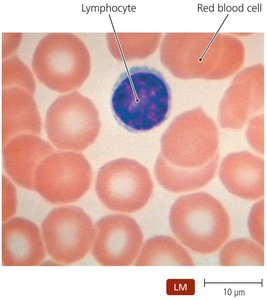
Both B and T cells circulate in the blood and lymphatic system, migrating to secondary lymphoid organs where they encounter antigens.
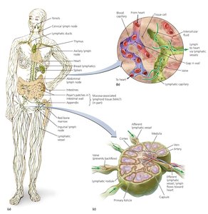
The Lymphatic System and Immune Surveillance
Tissues and Organs of the Lymphatic System
The lymphatic system is essential for immune surveillance and the initiation of adaptive immune responses. It consists of lymphatic vessels, lymphoid cells, tissues, and organs that screen the body for foreign molecules.
Lymphatic vessels: One-way system that returns lymph from tissues to the circulatory system.
Lymph: Fluid similar to blood plasma, derived from fluid leaked from blood vessels into tissues.
Primary lymphoid organs: Red bone marrow and thymus (sites of lymphocyte maturation).
Secondary lymphoid organs: Lymph nodes, spleen, tonsils, and mucosa-associated lymphoid tissue (MALT).
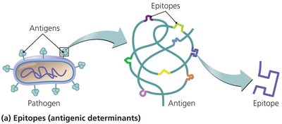
Antigens and Their Properties
Definition and Properties of Antigens
Antigens are molecules recognized as foreign by the immune system and capable of eliciting an immune response. The specific regions of antigens recognized by immune cells are called epitopes or antigenic determinants.
Large, complex macromolecules (proteins, polysaccharides) are the most effective antigens.
Antigens can be derived from microbes, viruses, fungi, protozoa, food, or dust.
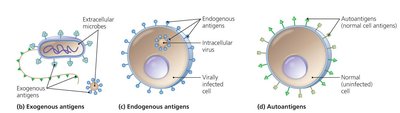
Types of Antigens
Exogenous antigens: Components from outside the cell, such as toxins, cell wall, membrane, flagella, and pili of microbes.
Endogenous antigens: Produced by microbes that replicate inside host cells.
Autoantigens: Derived from normal cellular processes; usually not targeted unless self-tolerance fails.
Major Histocompatibility Complex (MHC) and Antigen Presentation
Role of MHC Molecules
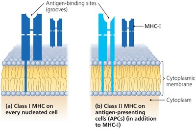
The major histocompatibility complex (MHC) consists of glycoproteins on cell membranes that present antigenic peptides to T cells. MHC molecules are crucial for distinguishing self from non-self and for initiating adaptive immune responses.
MHC class I: Present on all nucleated cells except red blood cells; present endogenous antigens.
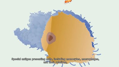
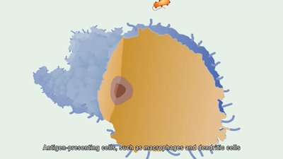
MHC class II: Present on antigen-presenting cells (APCs) such as macrophages, B cells, and dendritic cells; present exogenous antigens.
Antigen Processing and Presentation
Antigen processing involves breaking down antigens into smaller peptides that can be loaded onto MHC molecules for presentation to T cells. The process differs for endogenous and exogenous antigens.
Endogenous pathway: Processes antigens from within the cell (e.g., viral proteins) for presentation on MHC I.
Exogenous pathway: Processes antigens from outside the cell for presentation on MHC II.
T Lymphocytes (T Cells)
Development and Function
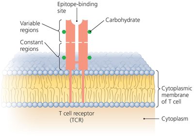
T cells are produced in the red bone marrow and mature in the thymus. They circulate in the blood and lymph, migrating to secondary lymphoid organs. T cells possess T cell receptors (TCRs) that recognize antigenic peptides presented by MHC molecules.
TCRs only bind to epitopes associated with MHC proteins.
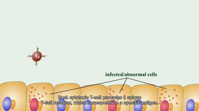
T cells primarily target cells harboring intracellular pathogens or abnormal proteins.
Types of T Lymphocytes
Cytotoxic T lymphocytes (Tc): Directly kill infected or abnormal cells.
Helper T lymphocytes (Th): Regulate B cells and cytotoxic T cells; include Th1 and Th2 subtypes.
Regulatory T lymphocytes (Tr): Suppress immune responses to prevent autoimmunity.
Lymphocyte | Site of Maturation | Surface Glycoproteins | Notable Secretions |
|---|---|---|---|
Helper T cell type 1 (Th1) | Thymus | CD4, TCR | Interleukin 2, IFN-γ |
Helper T cell type 2 (Th2) | Thymus | CD4, TCR | Interleukin 4, 5 |
Cytotoxic T cell (Tc) | Thymus | CD8, CD95L, TCR | Perforin, granzyme |
Regulatory T cell (Tr) | Thymus | CD4, CD25, TCR | Interleukin 10 |
B Lymphocytes (B Cells) and Antibodies
Development and Function
B cells are primarily found in the spleen, lymph nodes, and MALT, with a small percentage circulating in the blood. Their main function is the production and secretion of antibodies.
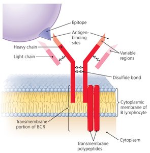
B Cell Receptor (BCR) Specificity
Each B cell expresses a unique B cell receptor (BCR) that binds to a specific epitope. The diversity of BCRs allows the immune system to recognize millions of different antigens.
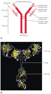
Antibody Structure and Function
Antibodies, or immunoglobulins, are secreted by activated B cells (plasma cells) and have antigen-binding sites identical to the BCR of the parent B cell. Antibodies function in several ways:
Activation of complement and inflammation
Neutralization of toxins and pathogens
Opsonization (enhancing phagocytosis)
Agglutination (clumping of antigens)
Antibody-dependent cellular cytotoxicity (ADCC)
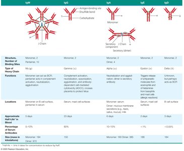
Classes of Antibodies
There are five main classes of antibodies, each with distinct functions and properties:
Class | Structure | Functions | Locations | Half-life |
|---|---|---|---|---|
IgM | Pentamer | First antibody produced; agglutination; complement activation | Blood, B cell surface | 5 days |
IgG | Monomer | Most abundant; crosses placenta; opsonization; neutralization | Blood, extracellular fluid | 23 days |
IgA | Dimer | Secretory antibody; protects mucosal surfaces | Secretions (tears, saliva, mucus) | 6 days |
IgE | Monomer | Allergic responses; defense against parasites | Bound to mast cells, basophils | 2 days |
IgD | Monomer | Function unclear; B cell receptor | B cell surface | 3 days |
Immune Response Cytokines
Types and Functions of Cytokines
Cytokines are soluble regulatory proteins that mediate communication between immune cells. They are secreted by various leukocytes and form a complex signaling network.
Interleukins (ILs): Signal among leukocytes.
Interferons (IFNs): Antiviral proteins; also act as cytokines.
Growth factors: Stimulate stem cell division.
Tumor necrosis factor (TNF): Induces inflammation or apoptosis.
Chemokines: Attract leukocytes to sites of infection.
Cytokine | Source | Target | Action |
|---|---|---|---|
Interleukin 2 (IL-2) | Th1, Tc cells | Tc cell | Cloning of Tc cell |
Interleukin 4 (IL-4) | Th2 cell | B cell | B cell differentiates into plasma cell |
Interleukin 12 (IL-12) | Dendritic cell | Th cell | Th cell differentiates into Th1 cell |
Gamma interferon (IFN-γ) | Th1 cell | Macrophage | Increases phagocytosis |
Tumor necrosis factor (TNF) | Macrophages, T cells | Body tissues | Triggers inflammation or apoptosis |
Cell-Mediated Immune Responses
Mechanisms and Functions
Cell-mediated immunity is primarily directed against intracellular pathogens (e.g., viruses, some bacteria and protozoa) and abnormal body cells (e.g., cancer cells). Cytotoxic T cells (Tc) are the main effectors, killing targets via two main pathways:
Perforin-granzyme pathway: Tc cells release perforin and granzyme to induce apoptosis in target cells.
CD95 pathway: Involves interaction with CD95 on target cells, triggering apoptosis.
Some activated T cells become memory T cells, which persist long-term and respond rapidly upon re-exposure to the same antigen.
Antibody (Humoral) Immune Responses
Activation and Function
Antibody-mediated (humoral) immunity targets exogenous pathogens and toxins. B cells are activated to proliferate and differentiate into plasma cells, which secrete antibodies specific to the encountered antigen.
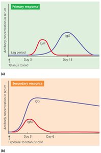
Immunological Memory
Memory B cells are produced during the primary immune response and persist in lymphoid tissues. Upon re-exposure to the same antigen, these cells rapidly initiate a secondary immune response, producing antibodies more quickly and in greater quantity than during the primary response.
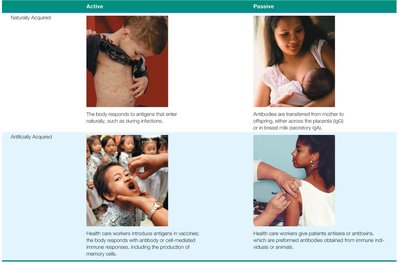
Types of Acquired Immunity
Classification of Acquired Immunity
Acquired immunity can be classified based on how it is obtained:
Naturally acquired: Immune response to antigens encountered in daily life (e.g., infection, maternal antibodies).
Artificially acquired: Immune response to antigens introduced via vaccination or administration of immune serum.
Both types can be active (host produces own antibodies) or passive (antibodies are transferred from another source).