 Back
BackBiochemical Identification of Bacteria: Key Differential Tests in Microbiology
Study Guide - Smart Notes
 Tailored notes based on your materials, expanded with key definitions, examples, and context.
Bacterial Biochemical Identification
Introduction
Biochemical tests are essential tools in microbiology for identifying and differentiating bacterial species, especially among Gram-negative rods such as the Enterobacteriaceae. These tests exploit differences in metabolic pathways, enzyme production, and fermentation abilities to distinguish between closely related organisms. Below are the major biochemical tests, their principles, procedures, and clinical relevance.
MR-VP Tests (Methyl Red and Voges–Proskauer)
Principle and Purpose
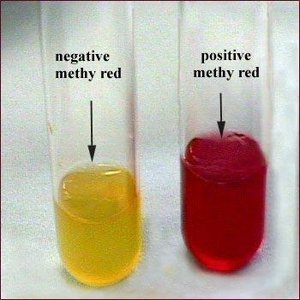
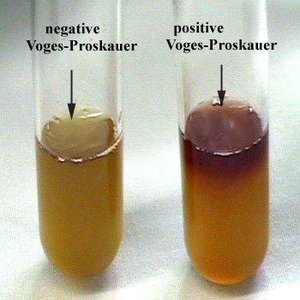
The Methyl Red (MR) and Voges–Proskauer (VP) tests are used together to differentiate bacteria based on their glucose fermentation pathways. The MR test detects strong acid production (mixed acid fermentation), while the VP test detects neutral end-products (acetoin, 2,3-butanediol) from glucose fermentation.
MR Test: Detects stable acid end-products. Positive result = red color (pH < 4.4); negative = yellow/orange (pH > 6).
VP Test: Detects acetoin. Positive result = red color after adding Barritt’s reagents; negative = no color change or copper tone.
Key organisms: Escherichia coli (MR+), Proteus vulgaris (MR+), Enterobacter and Klebsiella (VP+).
Procedure
Inoculate MR-VP broth with the test organism and incubate at 35–37°C for 48 hours.
Split the culture into two tubes.
For MR: Add methyl red indicator and observe color change.
For VP: Add Barritt’s reagents A and B, wait 15–30 minutes, and observe color change.
Interpretation Table
MR Result | VP Result | Interpretation | Example Organisms |
|---|---|---|---|
Red | No color | Mixed acid fermentation | E. coli, Proteus vulgaris |
Yellow/orange | Red | Butanediol fermentation | Enterobacter, Klebsiella |
Red | Red | Both pathways (rare) | Some E. coli strains |
Yellow | No color | No significant fermentation | Pseudomonas aeruginosa |
Indole Test
Principle and Purpose
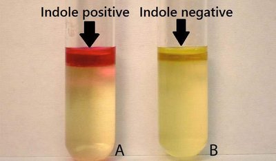
The Indole test detects the ability of bacteria to degrade tryptophan to indole using the enzyme tryptophanase. Indole reacts with Kovac’s reagent to produce a red or pink color at the top of the medium.
Positive: Red/pink layer (e.g., E. coli, Proteus vulgaris).
Negative: No color change/yellow (e.g., Klebsiella, Enterobacter).
Procedure
Inoculate tryptone broth with the test organism and incubate at 35–37°C for 24–48 hours.
Add a few drops of Kovac’s reagent.
Observe for a red/pink layer (positive) or no color change (negative).
Oxidase Test
Principle and Purpose
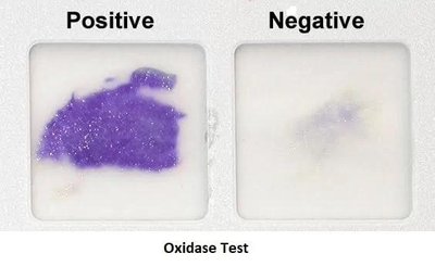
The Oxidase test identifies bacteria that produce cytochrome c oxidase, an enzyme of the electron transport chain. The reagent (TMPD) turns purple/blue when oxidized by cytochrome oxidase.
Positive: Dark purple/blue within 10–30 seconds (Pseudomonas aeruginosa, Neisseria spp.).
Negative: No color change or delayed color change (>60 sec) (all Enterobacteriaceae).
Procedure
Grow bacteria on non-selective medium.
Transfer a colony to filter paper or oxidase test strip.
Add oxidase reagent and observe color change within 10–30 seconds.
Nitrate Reduction Test
Principle and Purpose
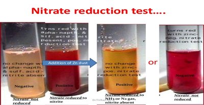
The Nitrate Reduction Test determines if bacteria can reduce nitrate (NO3−) to nitrite (NO2−) or further to nitrogen gas (N2), nitrous oxide (N2O), or ammonia (NH3). It is important for differentiating Gram-negative bacteria, especially Enterobacteriaceae.
Positive: Red after reagents A & B (nitrate → nitrite) or no color after zinc (nitrate reduced beyond nitrite).
Negative: Red only after zinc (nitrate not reduced).
Procedure
Inoculate nitrate broth and incubate at 35–37°C for 24–48 hours.
Add sulfanilic acid (A) and α-naphthylamine (B): red = positive.
If no color, add zinc powder: red = negative; no color = positive (reduced beyond nitrite).
Motility Test
Principle and Purpose
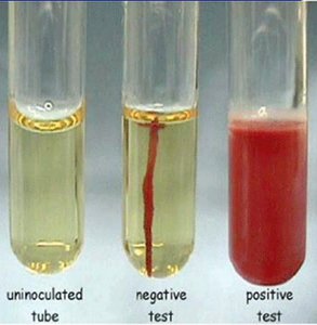
The Motility test determines whether bacteria can move through semi-solid agar, indicating the presence of flagella or other motility structures. Motile organisms diffuse away from the stab line, while non-motile organisms grow only along the stab line.
Motile: Diffuse/hazy growth (E. coli, Salmonella, Proteus).
Non-motile: Growth only on stab line (Klebsiella, Shigella).
Procedure
Inoculate semi-solid agar with a straight stab.
Incubate at 35–37°C for 24–48 hours.
Observe for diffuse or restricted growth.
Urease Hydrolysis Test
Principle and Purpose
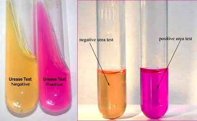
The Urease test detects the enzyme urease, which hydrolyzes urea to ammonia and carbon dioxide. Ammonia raises the pH, turning the phenol red indicator pink/magenta in alkaline conditions.
Positive: Pink/magenta (Proteus vulgaris, Proteus mirabilis, H. pylori).
Negative: Yellow/orange (E. coli, Salmonella, Shigella).
Procedure
Inoculate urea broth and incubate at 35–37°C.
Observe for color change to pink (positive) or yellow (negative).
Motility-Indole-Urease (MIU) Test
Principle and Purpose
The MIU test is a single-tube test that simultaneously assesses motility, indole production, and urease activity. It is efficient for presumptive identification of enteric bacteria.
Motility: Diffuse growth (positive) or growth only on stab line (negative).
Indole: Red/pink layer after Kovac’s reagent (positive).
Urease: Pink/magenta (positive), yellow/orange (negative).
Examples: Proteus vulgaris (M+, I+, U+); E. coli (M+, I+, U−); Klebsiella pneumoniae (M−, I−, U+).
Hydrogen Sulfide (H2S) Production Test
Principle and Purpose
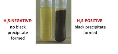
The H2S production test detects the ability of bacteria to produce hydrogen sulfide gas by reducing sulfur-containing compounds. H2S reacts with iron salts in the medium to form a black precipitate (FeS).
Positive: Black precipitate (Salmonella, Proteus).
Negative: No blackening (E. coli, Shigella, Klebsiella).
Procedure
Inoculate SIM, TSI, or KIA medium and incubate at 35–37°C for 18–24 hours.
Observe for black precipitate (positive) or no color change (negative).
Summary Table of Biochemical Tests
Test | Purpose | Principle | Positive Result | Negative Result | Example (Positive vs Negative) | Clinical Relevance |
|---|---|---|---|---|---|---|
Methyl Red (MR) | Detect mixed-acid glucose fermentation | Stable acids lower pH; methyl red detects low pH | Red (pH < 4.4) | Yellow/orange (pH > 6) | E. coli (+) vs Enterobacter (−) | Differentiates enteric Gram-negative rods |
Voges–Proskauer (VP) | Detect acetoin from butanediol pathway | Acetoin oxidized to red compounds | Red after 15–30 min | No color/copper | Enterobacter (+) vs E. coli (−) | Complements MR test |
Indole | Detect tryptophanase activity | Indole reacts with Kovac’s reagent | Red/pink ring | Yellow/no red | E. coli (+) vs Klebsiella (−) | Classic discriminator in enteric ID |
Oxidase | Detect cytochrome c oxidase | TMPD oxidized by enzyme | Purple/blue in 10–30 sec | No color | Pseudomonas (+) vs E. coli (−) | Separates non-enterics from Enterobacteriaceae |
Nitrate Reduction | Assess nitrate use in anaerobic respiration | Nitrate reduced to nitrite or beyond | Red after A+B or no color after zinc | Red only after zinc | Nitrate reducers (+) vs non-reducers (−) | Differentiates Gram-negative bacteria |
Motility | Determine motility | Motile bacteria move through agar | Diffuse/hazy growth | Growth on stab line | E. coli (+) vs Klebsiella (−) | Species-level differentiation |
Urease | Detect urease enzyme | Ammonia raises pH; phenol red turns pink | Pink/red | Yellow/orange | Proteus (+) vs E. coli (−) | Identifies urease producers (e.g., UTIs) |
MIU | Combined motility, indole, urease | Pattern-based | Variable | Variable | Proteus vulgaris: M+, I+, U+; E. coli: M+, I+, U− | Efficient presumptive ID |
H2S Production | Detect sulfur reduction | H2S reacts with iron salts | Black precipitate | No blackening | Salmonella (+) vs E. coli (−) | Distinguishes Salmonella/Proteus |
Medical Terms of the Week: Immune & Lymphatic System
Lympho-: Lymph or lymphatic system
Lymphaden-: Lymph nodes
Spleno-: Spleen
Immuno-: Immune system or immunity
Autoimmune: Immune system attacks the body’s own cells
Pathogen: Disease-causing microorganism
Antigen: Foreign substance triggering immune response
Antibody: Protein produced to neutralize antigens
Leukocyte: White blood cell
Inflammation: Tissue response to injury or infection (redness, heat, swelling, pain)
