 Back
BackCapsule Staining and Capsule-Forming Bacteria: Laboratory Study Guide
Study Guide - Smart Notes
 Tailored notes based on your materials, expanded with key definitions, examples, and context.
Capsule Staining: Principles and Procedure
Introduction to Capsule Staining
Capsule staining is a specialized microbiological technique used to visualize bacterial capsules, which are extracellular layers of gelatinous material that enhance pathogenicity and adherence. Capsules are typically composed of uncharged mucoid polysaccharides, though some species produce polypeptide capsules.
Capsule Function: Increases bacterial virulence and aids in adherence to surfaces.
Composition: Most capsules are polysaccharide-based; some, like Bacillus anthracis, are polypeptide.
Staining Challenge: Capsules are non-ionic and do not bind cationic stains such as Carbol Fuchsin.
Capsule Staining Procedure
The capsule stain involves mixing bacteria with albumin (egg white) to fix and provide a background, followed by staining with Carbol Fuchsin. The capsule appears as a clear halo around the stained cell.
Step 1: Label slide and add a loopful of distilled water (DiH2O).
Step 2: Add a loopful of Mayers Albumin.
Step 3: Add a scant amount of bacterial culture (e.g., Klebsiella, Enterobacter, Streptococcus) and mix well.
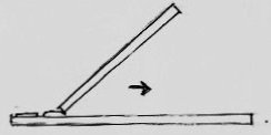
Step 4: Spread the mixture across the slide junction at a 45° angle to create a thin smear.
Step 5: Air dry the slide.
Step 6: Heat fix gently for 5 seconds (light smoke should be visible).
Step 7: Cool and flood with Ziehl-Neelsen Carbol Fuchsin for 10 seconds.
Step 8: Wash gently and air dry (do not blot).
Step 9: Observe under oil immersion lens.
Result: The cell and albumin background stain pinkish-red, while the capsule remains as a clear halo.
Capsule-Forming Bacteria: Characteristics and Clinical Relevance
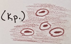
Klebsiella pneumoniae
Klebsiella pneumoniae is a Gram-negative, capsulated, straight rod bacterium found in various environments and clinical specimens.
Cell Morphology: 0.3–1.0 x 0.6–6 µm, occurs singly, in pairs, or short chains.
Metabolism: Facultative anaerobe; both respiratory and fermentative.
Habitat: Human feces, soil, water, grains, fruits, vegetables.
Pathogenicity: Opportunistic pathogen causing bacteremia, pneumonia, and UTIs.
Capsule: Enhances virulence and resistance to phagocytosis.
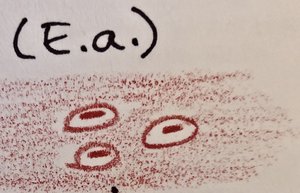
Enterobacter aerogenes
Enterobacter aerogenes is a Gram-negative, motile rod with peritrichous flagella, widely distributed in nature.
Cell Morphology: 0.6–1.0 x 1.2–3.0 µm.
Metabolism: Facultative anaerobe, chemoorganotrophic (respiratory and fermentative).
Habitat: Fresh water, soil, sewage, plants, vegetables, animal and human feces.
Pathogenicity: Opportunistic pathogen.
Capsule: Provides protection and increases pathogenic potential.
Streptococcus mutans
Streptococcus mutans is a Gram-positive, facultatively anaerobic coccus commonly found in the human oral cavity.
Cell Morphology: Round cells, often in chains.
Metabolism: Facultative anaerobe.
Habitat: Human oral cavity.
Pathogenicity: Major contributor to dental caries (tooth decay).
Capsule: Produces dextran, a sticky capsular material aiding in adherence to teeth.

Scientific and Clinical Importance of Capsule Staining
Role of Capsules in Pathogenicity
Capsules are critical for bacterial survival and virulence, providing resistance to host immune responses and facilitating colonization.
Immune Evasion: Capsules inhibit phagocytosis by host immune cells.
Adherence: Capsules enable bacteria to adhere to surfaces and tissues.
Virulence: Capsule presence correlates with increased disease severity.
Applications of Capsule Staining
Clinical Diagnosis: Identifies capsule-forming pathogens in patient samples.
Research: Studies bacterial virulence mechanisms and host-pathogen interactions.
Summary Table: Capsule-Forming Bacteria
Bacterium | Gram Reaction | Cell Shape | Capsule Composition | Pathogenicity |
|---|---|---|---|---|
Klebsiella pneumoniae | Negative | Rod | Polysaccharide | Bacteremia, pneumonia, UTIs |
Enterobacter aerogenes | Negative | Rod | Polysaccharide | Opportunistic infections |
Streptococcus mutans | Positive | Coccus | Dextran (polysaccharide) | Dental caries |
Key Terms and Definitions
Capsule: Extracellular, gelatinous layer surrounding some bacteria, composed of polysaccharides or polypeptides.
Facultative Anaerobe: Organism that can grow with or without oxygen.
Carbol Fuchsin: Basic dye used in capsule and acid-fast staining.
Dextran: Sticky polysaccharide produced by Streptococcus mutans.
Relevant Equations
Capsule Staining Principle
Staining Reaction: $\text{Capsule (non-ionic)} + \text{Carbol Fuchsin (cationic)} \rightarrow \text{No binding; capsule remains clear}$ $\text{Cell/Albumin} + \text{Carbol Fuchsin} \rightarrow \text{Pinkish-red staining}$
Conclusion
Capsule staining is a vital technique in microbiology for identifying and studying capsule-forming bacteria, which are often significant pathogens. Understanding the procedure and interpretation of capsule stains aids in clinical diagnosis and research into bacterial virulence.
