 Back
BackCharacterizing and Classifying Viruses, Viroids, and Prions: Study Notes
Study Guide - Smart Notes
 Tailored notes based on your materials, expanded with key definitions, examples, and context.
Characterizing and Classifying Viruses, Viroids, and Prions
Characteristics of Viruses
Viruses are unique infectious agents that differ fundamentally from cellular life forms. They are responsible for numerous diseases across all domains of life and possess distinctive structural and functional properties.
Definition: Viruses are minuscule, acellular entities containing either DNA or RNA as their genetic material, but never both.
Infectious Nature: They infect humans, animals, plants, fungi, protists, and bacteria.
Metabolic Inactivity: Viruses cannot carry out metabolic pathways, grow, or respond to their environment independently.
Replication: They rely on host cell machinery for reproduction, recruiting the cell's metabolic pathways to increase their numbers.
Structure: Viruses lack cytoplasmic membrane, cytosol, and organelles.
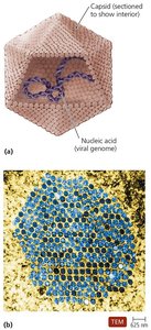
States: Exist in extracellular (virion) and intracellular (nucleic acid) states.
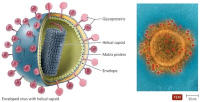
Extracellular State (Virion)
Virion: The complete virus particle, consisting of a nucleic acid surrounded by a protein coat (capsid).
Nucleocapsid: The combination of nucleic acid and capsid.
Envelope: Some virions possess a phospholipid envelope derived from the host cell.
Function: The outermost layer provides protection and recognition sites for host cells.
Intracellular State
Capsid Removal: Upon entry into the host cell, the capsid is removed.
Nucleic Acid: The virus exists as naked nucleic acid within the host cell.
Genetic Material of Viruses
Viruses exhibit remarkable diversity in their genetic material, which is a primary basis for their classification.
Types: May be DNA or RNA, but never both.
Forms: Can be double-stranded (dsDNA, dsRNA) or single-stranded (ssDNA, ssRNA).
Structure: Genomes may be linear and segmented or single and circular.
Size: Viral genomes are much smaller than those of cells.
Hosts of Viruses
Viruses are highly specific in their host range, determined by molecular interactions between viral and host cell surface proteins.
Specificity: Most viruses infect only particular host cells due to affinity of viral surface proteins for complementary host proteins.
Generalists: Some viruses can infect multiple cell types or hosts.
Universality: All types of organisms are susceptible to some virus.
Capsid Morphology
The capsid is a protein shell that encases the viral genome, providing protection and facilitating attachment to host cells.
Capsomeres: Capsids are composed of protein subunits called capsomeres, which may be made of single or multiple types of proteins.
Function: Protection and attachment to host cells.
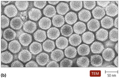
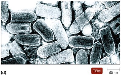
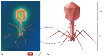
Viral Shapes
Viruses are classified based on the shape of their virions, which can be observed under electron microscopy.
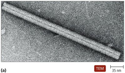
Helical: Rod-shaped, with nucleic acid spiraling within a cylindrical capsid.
Polyhedral: Spherical or many-sided, often icosahedral.
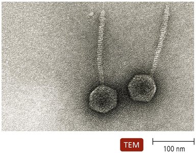
Complex: More elaborate structures, such as bacteriophages with head and tail regions.
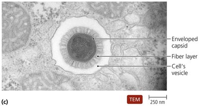
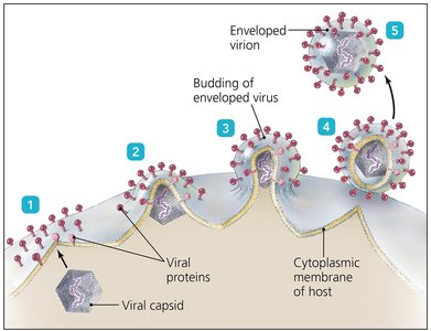
The Viral Envelope
Some viruses possess an envelope, which is acquired from the host cell during replication or release.
Composition: Phospholipid bilayer and proteins, including virally coded glycoproteins (spikes).
Function: Envelope proteins and glycoproteins play a role in host recognition.
Fragility: Enveloped viruses are more fragile than naked viruses.
Classification of Viruses
Viruses are classified based on several criteria, though their relationships are not fully understood.
Criteria: Type of nucleic acid, presence of envelope, shape, and size.
Taxonomy: Viral genera are organized into families.
Viral Replication
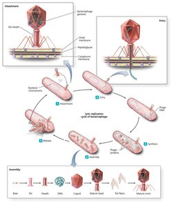
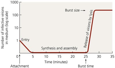
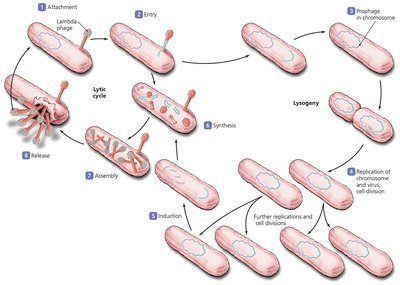
Lytic Replication Cycle
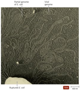
The lytic cycle is a process by which viruses replicate, resulting in the destruction of the host cell.
Attachment: Virus attaches to host cell.
Entry: Viral genome enters host cell.
Synthesis: Host cell machinery synthesizes viral components.
Assembly: New virions are assembled.
Release: Host cell lyses, releasing new virions.
Lysogenic Replication Cycle
Some bacteriophages undergo a lysogenic cycle, where the viral genome integrates into the host chromosome and replicates passively.
Temperate Phages: Can enter lysogenic cycle.
Prophage: Inactive phage DNA integrated into host genome.
Lysogenic Conversion: Phages may carry genes that alter the phenotype of the bacterium.
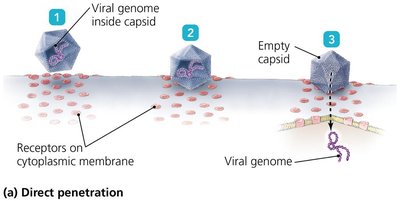
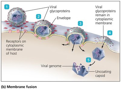
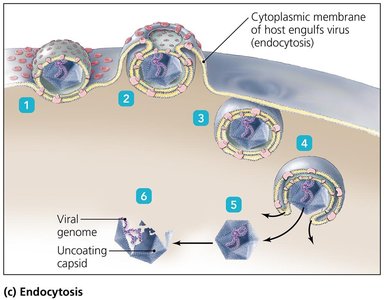
Replication of Animal Viruses
Animal viruses replicate using similar pathways to bacteriophages, with differences due to the presence of envelopes and the eukaryotic nature of host cells.
Attachment: Mediated by glycoprotein spikes or other molecules.
Entry Mechanisms: Direct penetration, membrane fusion, or endocytosis.
Synthesis: DNA viruses often replicate in the nucleus; RNA viruses in the cytoplasm.
Retroviruses: Use reverse transcriptase to produce DNA from RNA template.
Assembly and Release: DNA viruses assemble in nucleus; RNA viruses in cytoplasm. Enveloped viruses bud from host membrane; naked viruses released by exocytosis or lysis.
Latency of Animal Viruses
Latent Viruses: Remain dormant in host cells as proviruses.
Duration: Latency may last years with no viral activity.
Permanence: Incorporation of provirus into host DNA is permanent.
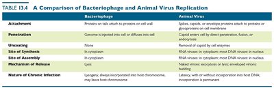
Comparison of Bacteriophage and Animal Virus Replication
The replication cycles of bacteriophages and animal viruses share similarities but also exhibit distinct differences.
Step | Bacteriophage | Animal Virus |
|---|---|---|
Attachment | Proteins on tail attach to proteins on cell wall | Spikes, capsid, or envelope proteins attach to proteins or glycoproteins on cell membrane |
Penetration | Genome is injected into cell or diffuses into cell | Capsid enters cell by direct penetration, fusion, or endocytosis |
Uncoating | None | Removed by cell enzymes |
Site of Synthesis | In cytoplasm | RNA viruses in cytoplasm; most DNA viruses in nucleus |
Site of Assembly | In cytoplasm | RNA viruses in cytoplasm; most DNA viruses in nucleus |
Mechanism of Release | Lysis | Enveloped viruses bud; naked viruses exocytosis or lysis |
Nature of Chronic Infection | Lysogeny; always incorporated into host chromosome, may leave host chromosome | Latency, with or without incorporation into host DNA; incorporation is permanent |
The Role of Viruses in Cancer
Oncogenes and Cancer Induction
Viruses can contribute to cancer development by affecting cellular genes that regulate growth and division.
Protooncogenes: Genes that promote cell growth and division.
Oncogenes: Uncontrolled activation can lead to cancer.
Environmental Factors: Ultraviolet light, radiation, carcinogens, and viruses can activate oncogenes.
Mechanisms: Some viruses carry oncogenes, promote host oncogenes, or interfere with tumor repression.
Examples: Burkitt’s lymphoma, Hodgkin’s disease, Kaposi’s sarcoma, cervical cancer.
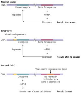
Summary Table: Virus-Induced Cancer Mechanisms
Viruses cause 20–25% of human cancers.
Mechanisms include carrying oncogenes, promoting host oncogenes, and interfering with tumor repression.
Example: Human papillomavirus (HPV) is known to cause cervical cancer by interfering with tumor suppressor genes.
----------------------------------------
