 Back
BackCharacterizing and Classifying Viruses, Viroids, and Prions
Study Guide - Smart Notes
 Tailored notes based on your materials, expanded with key definitions, examples, and context.
Characterizing and Classifying Viruses, Viroids, and Prions
Characteristics of Viruses
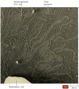
Viruses are acellular infectious agents that contain either DNA or RNA as their genetic material. They are obligate intracellular parasites, meaning they require a host cell to replicate and cannot carry out metabolic processes independently.
Acellular: Lack cellular structure, cytoplasmic membrane, cytosol, and organelles.
Genetic Material: May be DNA or RNA, single- or double-stranded, linear, circular, or segmented.
Replication: Cannot reproduce independently; must hijack host cell machinery.
States: Exist in extracellular (virion) and intracellular (nucleic acid) states.
Genetic Material of Viruses
The primary method for classifying viruses is based on their genetic material. Viral genomes are much smaller than those of cells and can be highly variable in structure.
Types: dsDNA, ssDNA, dsRNA, or ssRNA.
Structure: Linear, circular, or segmented.
Examples: Influenzavirus (segmented RNA), Poliovirus (single RNA).
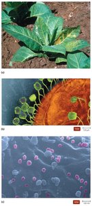
Hosts of Viruses
Viruses exhibit host specificity, often infecting only particular species or cell types due to the affinity between viral surface proteins and host cell receptors.
Specific viruses: Infect only certain cells (e.g., HIV infects human T cells).
Generalist viruses: Infect multiple hosts or cell types (e.g., West Nile virus).
All life forms: All organisms are susceptible to some form of virus.
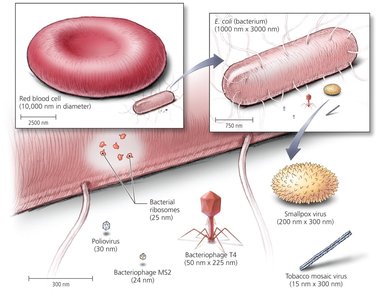
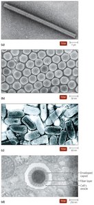
Size and Morphology of Virions
Virions vary greatly in size and shape, typically much smaller than bacterial cells. Their structure is essential for protection and host recognition.
Size: Range from 10 nm to over 500 nm.
Comparison: Viruses are much smaller than bacteria and eukaryotic cells.
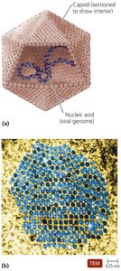
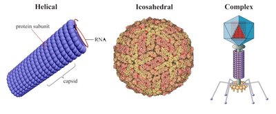
Capsid Morphology and Viral Shapes
The capsid is a protein shell that encases the viral genome, composed of subunits called capsomeres. Viruses are classified by their capsid shape into three main types:
Helical: Rod-shaped, with the genome coiled inside.
Polyhedral (Icosahedral): Spherical with 20 faces.
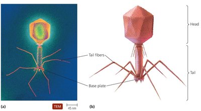
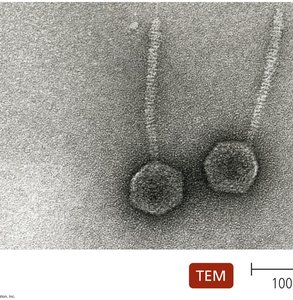
Complex: More intricate structures, often seen in bacteriophages.
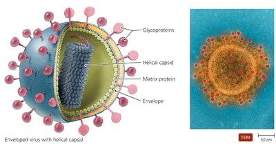
The Viral Envelope
Some viruses possess an envelope derived from the host cell membrane, which contains viral glycoproteins essential for host recognition and immune evasion.
Composition: Phospholipid bilayer and proteins (some virally encoded as spikes).
Function: Protects from the immune system but makes the virus more fragile outside the host.
Naked viruses: Lack an envelope, are more stable in the environment but more susceptible to immune defenses.
Classification of Viruses
Viruses are classified based on their nucleic acid type, presence of an envelope, shape, and size. Viral taxonomy is less developed than for cellular organisms, with genera grouped into families.
DNA Viruses: Families include Poxviridae, Herpesviridae, Papillomaviridae, etc.
RNA Viruses: Families include Picornaviridae, Flaviviridae, Retroviridae, etc.
Family names: Often derived from key characteristics or important members.
Viral Replication Cycles
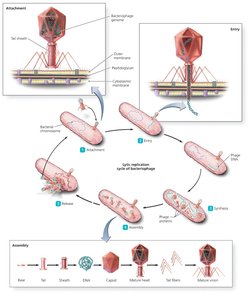
Viruses replicate by commandeering host cell machinery. The two main cycles in bacteriophages are the lytic and lysogenic cycles.
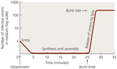
Lytic Replication Cycle
Attachment: Virus binds to host cell receptors.
Entry: Viral genome enters the host cell, often via injection.
Synthesis: Host machinery synthesizes viral components.
Assembly: New virions are assembled.
Release: Host cell lyses, releasing new virions.
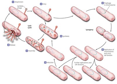
Lysogenic Replication Cycle
Integration: Viral genome integrates into host DNA as a prophage.
Replication: Host cell divides, copying the prophage.
Induction: Environmental triggers can reactivate the prophage, entering the lytic cycle.
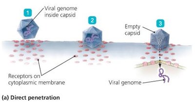
Replication of Animal Viruses
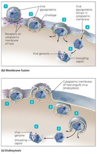
Animal viruses follow similar steps but differ due to the presence of envelopes and the eukaryotic nature of host cells. Entry can occur via direct penetration, membrane fusion, or endocytosis.
Attachment: Mediated by glycoprotein spikes or other molecules.
Entry: Three mechanisms: direct penetration, membrane fusion, endocytosis.
Uncoating: Removal of capsid to release viral genome.
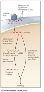
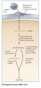
Synthesis of Animal Viruses
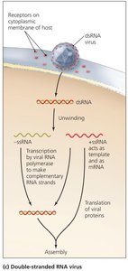
The strategy for genome replication and mRNA synthesis depends on the type of viral nucleic acid.
dsDNA viruses: Replicate in the nucleus, proteins made in cytoplasm.
ssDNA viruses: Form dsDNA intermediates for replication.
+ssRNA viruses: Genome acts as mRNA.
Retroviruses: Use reverse transcriptase to make DNA from RNA.
-ssRNA viruses: Require RNA-dependent RNA transcriptase to make +ssRNA.
dsRNA viruses: Each strand serves as a template for its complement.
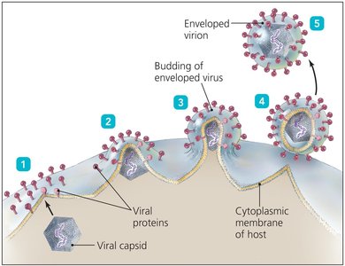
Assembly and Release of Animal Viruses
Assembly: DNA viruses assemble in the nucleus; RNA viruses in the cytoplasm.
Release: Enveloped viruses bud from the cell membrane; naked viruses are released by lysis or exocytosis.
Latency of Animal Viruses
Latent viruses (proviruses): Remain dormant in host cells, sometimes permanently incorporated into host DNA (e.g., HIV, herpesviruses).
The Role of Viruses in Cancer
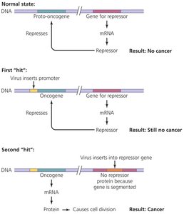
Some viruses can induce cancer by disrupting normal cell cycle regulation, often by carrying oncogenes or interfering with tumor suppressor genes.
Neoplasia: Uncontrolled cell division resulting in tumors.
Oncogene Theory: Multiple mutations or 'hits' are required for cancer development.
Examples: EBV (Burkitt’s lymphoma), HPV (cervical cancer).
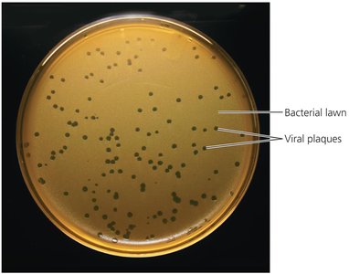
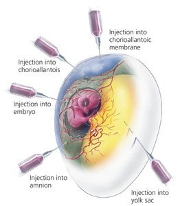
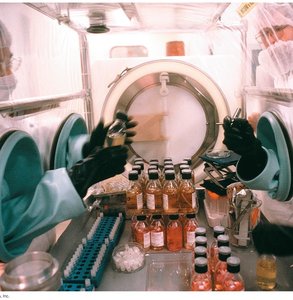
Culturing Viruses in the Laboratory
Viruses require living cells for replication and are cultured using mature organisms, embryonated eggs, or cell cultures.
Mature organisms: Bacteria (for phages), plants, or animals.
Embryonated eggs: Common for vaccine production.
Cell cultures: Diploid (finite) or continuous (immortal, e.g., HeLa cells).
Viroids and Prions
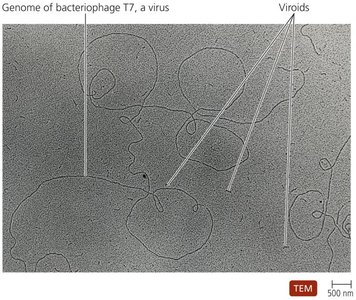
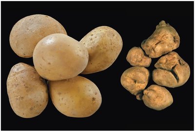
Viroids
Viroids are small, circular, single-stranded RNA molecules that infect plants. They lack a protein coat and do not encode proteins, causing disease by interfering with host gene expression.
Viroidlike agents: Infect some fungi.
Prions
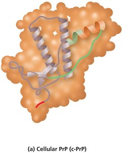
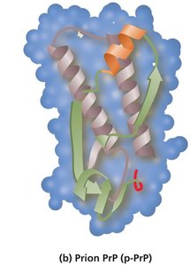
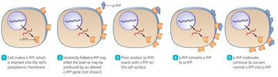
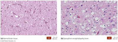
Prions are infectious proteins that cause neurodegenerative diseases by inducing misfolding of normal cellular proteins (PrP) into the pathogenic form.
Cellular PrP (c-PrP): Normal, α-helix-rich structure.
Prion PrP (p-PrP): Disease-causing, β-sheet-rich structure.
Diseases: BSE (mad cow disease), CJD, scrapie, kuru, chronic wasting disease.
Transmission: Inherited, sporadic, or infectious routes.
Resistant to sterilization: Only destroyed by incineration or autoclaving in strong alkali.
Comparison Table: Bacteria, Viruses, Viroids, and Prions
Bacteria | Viruses | Viroids | Prions | |
|---|---|---|---|---|
Width | 200–2000 nm | 10–400 nm | 2 nm | 5 nm |
Length | 200–550,000 nm | 20–800 nm | 40–130 nm | 5 nm |
Nucleic Acid | DNA and RNA | DNA or RNA | RNA only | None |
Protein | Present | Present | Absent | Present (PrP) |
Cellular | Yes | No | No | No |
Cytoplasmic Membrane | Present | Absent (some have envelope) | Absent | Absent |
Functional Ribosomes | Present | Absent | Absent | Absent |
Growth | Present | Absent | Absent | Absent |
Self-Replicating | Yes | No | No | No; transforms PrP protein already present |
Responsiveness | Present | Some bacteriophages respond to host cell | Absent | Absent |
Metabolism | Present | Absent | Absent | Absent |
