 Back
BackEssential Study Notes: Introduction to Microbiology, Microscopy, Cell Structure, Viruses, and Microbial Growth
Study Guide - Smart Notes
 Tailored notes based on your materials, expanded with key definitions, examples, and context.
CH 1: The Microbial World and You
Introduction to Microorganisms
Microorganisms are organisms too small to be seen with the naked eye. They play crucial roles in the environment, industry, and health. The study of these organisms is fundamental to understanding life processes and disease.
Types of Microorganisms: Bacteria, Fungi, Protozoa, Microscopic Algae, Viruses, Prions
Roles of Microbes:
Recycle nutrients and breakdown organic matter
Decompose organic waste
Incorporate nitrogen gas from the air
Produce food (e.g., fermentation, photosynthesis)
Used in manufacturing (e.g., cellulose, insulin)
Some are pathogens causing disease or food spoilage
Microbiome: The community of microbes living on and in the human body; the human body contains about 3 billion body cells and harbors approximately 40 trillion bacterial cells.
Scientific Nomenclature
Microorganisms are named using a binomial system established by Carolus Linnaeus in 1735. The first name is the genus and the second is the species.
Example: Escherichia coli
Scientific names provide clues about the organism's characteristics or habitat.
Classification of Microorganisms
Carl Woese (1978) proposed three domains based on cellular organization:
Bacteria
Archaea
Eukarya
Applications of Microbiology
Sustainable energy production
Recycling vital elements: Bacteria convert carbon, oxygen, sulfur, and phosphorus into usable forms.
Bioremediation: Use of microbes to degrade pollutants (e.g., oil spills, sewage detoxification).
Biotechnology: Use of microbes to produce food, chemicals, and energy.
Recombinant DNA technology: Genetically modified organisms (GMOs) for crop protection and medicine.
CH 3: Observing Microorganisms Through a Microscope
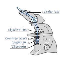
Microscope Structure and Function
Microscopes are essential tools for observing microorganisms. Understanding their parts and functions is crucial for effective use.
Ocular lens: The eyepiece through which the specimen is viewed.
Objective lens: Provides primary magnification.
Condenser lenses: Focus light onto the specimen.
Diaphragm: Regulates the amount of light reaching the specimen.
Illuminator: Light source for the microscope.
Preparing Smears and Staining
Staining enhances contrast in microscopic specimens, allowing for better visualization of cellular structures.
Smear: A thin film of sample spread over a slide.
Fixing: Adheres cells to the slide, usually by heat or chemicals, before staining.
Staining Types:
Simple stain: Uses one dye (e.g., methylene blue, crystal violet) to reveal shape and structure.
Differential stain: Distinguishes between types of bacteria (e.g., Gram stain, Acid-fast stain).
Special stain: Highlights specific structures (e.g., capsule, flagella, endospore stains).
Mordant: Substance used to hold stain or coat the specimen.
Gram Stain
The Gram stain differentiates bacteria based on cell wall structure:
Gram-positive: Thick peptidoglycan wall; stains purple.
Gram-negative: Thin wall; stains red/pink.
Counterstain: Applied at the end to visualize Gram-negative cells.
Acid-Fast Stain
Used to identify bacteria with waxy cell walls (e.g., Mycobacterium species). Acid-fast cells appear red.
CH 4: Functional Anatomy of Prokaryotic and Eukaryotic Cells
Prokaryotic vs. Eukaryotic Cells
Cells are classified as prokaryotic or eukaryotic based on structural differences.
Prokaryotes: Simpler, smaller, no nucleus, no membrane-bound organelles. Examples: Bacteria, Archaea.
Eukaryotes: More complex, larger, have a true nucleus and membrane-bound organelles. Examples: Fungi, Protozoa, Algae, Helminths.
Bacterial Shapes and Arrangements
Coccus: Spherical; may form chains, clusters, or cubes.
Bacillus: Rod-shaped; may be single or in chains.
Spiral: Spiral-shaped bacteria.
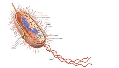
Structure of Prokaryotic Cells
Prokaryotic cells have specialized structures for survival and function.
Cell envelope: Complex boundary with multiple layers.
Pili: Used for DNA exchange between cells.
Flagella: Used for motility.
Fimbriae: Protein structures for attachment.
Glycocalyx: Capsule or slime layer for protection and attachment.
Plasma membrane: Contains enzymes for ATP production.
Cytoplasm: Thick substance inside the cell.
Ribosomes: 70S type; site of protein synthesis.
Nucleoid: Region containing the bacterial chromosome.
Endospore: Resistant, dormant structure for survival in harsh conditions.
Structure of Eukaryotic Cells
Nucleus: Double membrane, contains DNA and histones.
Endoplasmic Reticulum (ER): Rough ER (with ribosomes) synthesizes proteins; Smooth ER synthesizes fats and hormones.
Mitochondria: Site of ATP production; double membrane.
Ribosomes: 80S type (larger); 70S in plant organelles.
Flagella/Cilia: Used for movement; composed of microtubules.
Cell wall: Present in plants and algae; composed of cellulose or other polysaccharides.
CH 13: Viruses, Viroids, and Prions
Features of Viruses
Viruses are acellular entities that require a host cell to replicate. They are much smaller than bacteria.
Genetic Material: DNA or RNA (never both), single or double-stranded.
Capsid: Protein coat made of capsomeres; protects nucleic acid and aids in attachment.
Envelope: Lipid, protein, and carbohydrate coating; may carry host-derived molecules.
Spikes: Glycoprotein projections for host cell recognition and attachment.
Virion: Complete, infectious viral particle.
Viral Replication
Adsorption: Virus attaches to host cell using spikes.
Penetration: Virus enters the cell via vesicle or fusion.
Uncoating: Capsid is removed, releasing genetic material.
Synthesis: Host cell synthesizes viral components.
Assembly: New virions are assembled; spike proteins inserted into host membrane.
Release: New virions exit the host cell.
Antiviral drugs often target specific steps in this cycle.
Prions and Viroids
Prions: Infectious proteins causing neurodegenerative diseases (e.g., Creutzfeldt-Jakob disease, mad cow disease).
Viroids: Small, naked RNA molecules infecting plants, causing stunted growth (e.g., in potatoes, tomatoes).
CH 6: Microbial Growth
Environmental Factors Affecting Microbial Growth
Microbial growth is influenced by physical and chemical factors.
Physical factors: Temperature, pH, osmotic pressure.
Chemical factors: Sources of carbon, nitrogen, sulfur, phosphorus, oxygen.
Temperature Requirements
Psychrophiles: Cold-loving; grow below 15°C.
Psychrotrophs: Grow between 15°C and 30°C.
Mesophiles: Grow best between 30°C and 40°C.
Thermoduric: Survive short exposure to high temperatures.
Thermophiles: Heat-loving; grow between 45°C and 60°C.
Extreme thermophiles: Grow between 80°C and 121°C.
Oxygen Requirements
Obligate aerobes: Require oxygen (growth at top of tube).
Facultative anaerobes: Can grow with or without oxygen (growth throughout, mostly at top).
Microaerophiles: Require low oxygen levels.
Obligate anaerobes: Cannot tolerate oxygen (growth at bottom).
Aerotolerant anaerobes: Can survive in oxygen but do not use it.
Oxygen requirements can be determined using reducing media (e.g., thioglycolate broth).
pH and Osmotic Pressure
Most bacteria: Grow best between pH 6.5 and 7.5.
Molds and yeasts: Grow between pH 5 and 6.
Acidophiles: Grow in acidic environments.
Osmotic pressure: High salt environments cause plasmolysis; extreme halophiles require high salt, facultative halophiles tolerate it.
Culture Media
General-purpose media: Nutrient agar, broth, brain heart infusion, trypticase soy agar.
Enriched media: Contains complex organic substances (e.g., blood agar, chocolate agar).
Selective-differential media: Allows growth of some organisms while inhibiting others (e.g., Mannitol Salt Agar, Eosin Methylene Blue, MacConkey's Agar).
Bacterial Growth Curve
Lag phase: Adaptation, no increase in number.
Exponential (log) phase: Rapid cell division.
Stationary phase: Growth rate slows; nutrients deplete, waste accumulates.
Death phase: Cells die faster than they divide.
