 Back
BackFoundations of Microbiology: Microscopy, Staining, and Pioneers
Study Guide - Smart Notes
 Tailored notes based on your materials, expanded with key definitions, examples, and context.
The Microbial World and the Birth of Microbiology
Discovery of Microorganisms
The field of microbiology began with the discovery and observation of microorganisms, which are organisms too small to be seen with the naked eye. Early pioneers used primitive microscopes to reveal this hidden world, fundamentally changing our understanding of biology and disease.
Robert Hooke (1665): First to describe microbes and coined the term "cell" after observing fungal fruiting bodies.
Antonie van Leeuwenhoek (1676): First to observe and describe bacteria, which he called "wee animalcules." He constructed simple microscopes with remarkable magnifying power.
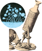
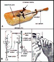
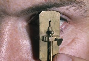
Example: Leeuwenhoek's observations of pond water revealed a diversity of microscopic life, including bacteria and protozoa.
Microscopy: Tools for Seeing the Invisible
Principles of Light Microscopy
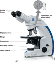
Light microscopy uses visible light and a series of lenses to magnify small objects. It is the foundational tool for observing microbial cells.
Bright-field microscopy: The standard form, where light passes through the specimen. Key components include the condenser, objective lens (10–100×), and ocular lens (10–30×).
Total magnification: Calculated as the product of the objective and ocular lens magnifications.
Resolution: The ability to distinguish two close objects as separate. The practical limit for light microscopy is about 0.2 µm.
Magnification vs. Resolution: Magnification makes objects appear larger, but resolution determines clarity and detail.
Resolution and the Abbe Equation
The resolving power of a microscope is described by the Abbe equation, which relates the minimal resolvable distance to the wavelength of light and the numerical aperture of the lens system.
Index of refraction: Affects resolution; oil immersion (n ≈ 1.515) is used to increase numerical aperture and improve resolution.
Abbe Equation:
Where:
= minimal distance to distinguish two points
= wavelength of light
= numerical aperture
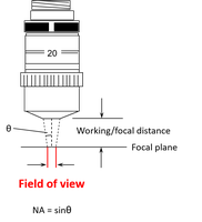
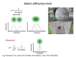
Example: Using oil immersion increases , thus decreasing and improving resolution.
Improving Contrast: Staining Techniques
Microbial cells are often transparent and require staining to increase contrast for observation under bright-field microscopy.
Basic dyes: Positively charged, bind to negatively charged cell components (e.g., nucleic acids, cell surfaces). Examples include methylene blue, crystal violet, and safranin.
Simple stain: Involves drying, heat-fixing, and staining cells to visualize their shape and arrangement.
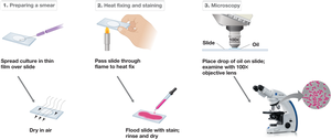
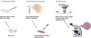
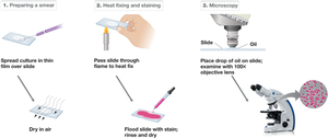
Example: Heat fixing kills cells and adheres them to the slide, while staining reveals cell morphology.
Differential Staining: The Gram Stain
The Gram stain is a differential staining technique that distinguishes bacteria based on cell wall structure.
Procedure: Involves sequential application of crystal violet, iodine, alcohol (decolorizer), and safranin.
Results: Gram-positive cells retain crystal violet (purple); Gram-negative cells are decolorized and counterstained pink/red by safranin.
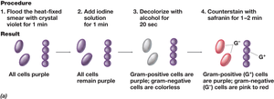
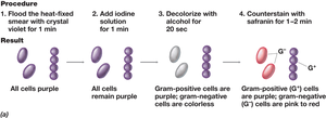
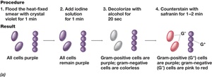
Example: Staphylococcus aureus (Gram-positive) appears purple, while Escherichia coli (Gram-negative) appears pink after Gram staining.
Pioneers of Microbiology
Louis Pasteur (1822–1895)
Pasteur's experiments established the role of microbes in fermentation and disease, and he developed methods for sterilization and vaccination.
Disproved spontaneous generation with swan-neck flask experiments.
Developed pasteurization and vaccines for anthrax, fowl cholera, and rabies.
Example: Pasteur's swan-neck flask experiment showed that sterilized broth remained free of microbes unless exposed to airborne contaminants.
Robert Koch (1843–1910)
Koch developed solid media for culturing microbes and established criteria (Koch's postulates) for linking specific microbes to specific diseases.
Identified causative agents of anthrax, tuberculosis, and cholera.
Formulated Koch's postulates for disease causation:
Step | Description |
|---|---|
1 | Pathogen must be found in all cases of the disease |
2 | Pathogen must be isolated in pure culture |
3 | Pathogen must cause disease when introduced into a healthy host |
4 | Pathogen must be re-isolated from the experimentally infected host |
Example: Koch used these postulates to demonstrate that Bacillus anthracis causes anthrax.
Sergei Winogradsky (1856–1953)
Winogradsky linked specific bacteria to biogeochemical cycles and discovered chemolithotrophy and autotrophy.
Demonstrated microbial roles in nitrogen and sulfur cycles.
Identified Clostridium pasteurianum as the first anaerobic nitrogen-fixing microbe.
Developed the Winogradsky column to study microbial communities.
Example: The Winogradsky column demonstrates the stratification of microbial metabolism in a gradient of nutrients and oxygen.
Martinus Beijerinck (1851–1931)
Beijerinck pioneered enrichment culture techniques and was the first to observe viruses, launching the field of virology.
Isolated Azotobacter chroococcum, the first aerobic nitrogen-fixing bacterium.
Used the Chamberland filter to show that infectious agents smaller than bacteria (viruses) could pass through and cause disease.
Example: Beijerinck's work with tobacco mosaic virus demonstrated that the infectious agent was not a bacterium but a new type of pathogen, later known as a virus.
Other Key Contributors
Ignaz Semmelweis: Advocated handwashing to reduce infection in hospitals.
Joseph Lister: Introduced aseptic techniques in surgery.
Albert Jan Kluyver: Proposed the unity of biochemistry across all life forms.
Summary Table: Types of Microscopy
Type | Principle | Resolution | Application |
|---|---|---|---|
Bright-field | Light passes through specimen | ~0.2 µm | General cell observation |
Phase-contrast | Contrast from refractive index differences | ~0.2 µm | Live, unstained cells |
Fluorescence | Fluorescent dyes or proteins | ~0.2 µm | Specific structures, tagged molecules |
Electron (TEM/SEM) | Electron beams | ~0.2 nm | Ultrastructure, viruses |
Additional info: Modern microbiology continues to build on these foundational discoveries, using advanced microscopy and molecular techniques to explore microbial diversity, physiology, and their roles in health and the environment.
