 Back
BackFunctional Anatomy of Prokaryotic and Eukaryotic Cells: Structure and Function
Study Guide - Smart Notes
 Tailored notes based on your materials, expanded with key definitions, examples, and context.
Comparing Prokaryotic and Eukaryotic Cells
Basic Differences
Prokaryotic and eukaryotic cells differ fundamentally in their structure and organization. Understanding these differences is essential for studying microbial anatomy and physiology.
Prokaryotes: "Pre-nucleus"; lack a true nucleus and membrane-bound organelles. DNA is typically a single circular chromosome located in the nucleoid region.
Eukaryotes: "True nucleus"; possess a nucleus enclosed by a nuclear membrane and various membrane-bound organelles. DNA is organized into paired chromosomes.
Cell Division: Prokaryotes divide by binary fission; eukaryotes divide by mitosis.
Cell Walls: Bacterial prokaryotes have peptidoglycan cell walls; archaea have pseudomurein; eukaryotic cell walls (when present) are made of polysaccharides.
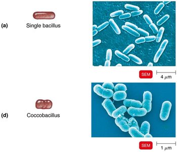
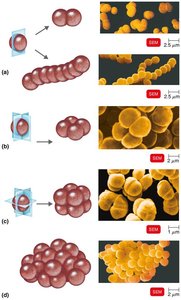
Size, Shape, and Arrangement of Bacteria
Bacterial Morphology
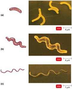
Bacteria exhibit a variety of shapes and arrangements, which are important for identification and classification.
Bacillus: Rod-shaped
Coccus: Spherical-shaped
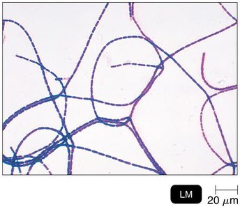
Spiral Forms: Includes vibrio (comma-shaped), spirillum (rigid spiral), and spirochete (flexible spiral)
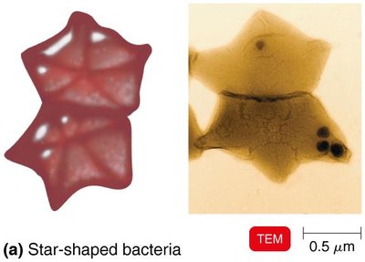
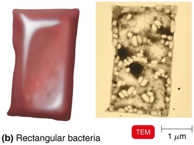
Other Shapes: Star-shaped and rectangular bacteria are less common.
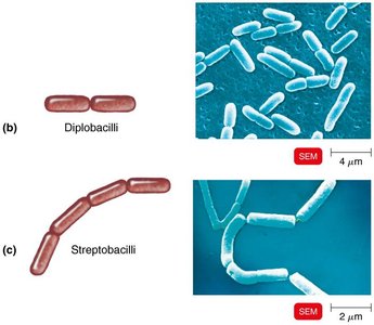
Arrangements:
Pairs: Diplococci, diplobacilli
Chains: Streptococci, streptobacilli
Clusters: Staphylococci
Groups of Four: Tetrads
Cubelike Groups of Eight: Sarcinae
The Structures of a Prokaryotic Cell
General Structure
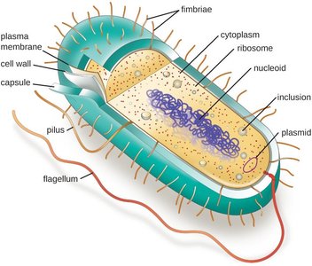
Prokaryotic cells have several key structures that contribute to their survival, pathogenicity, and classification.
Capsule/Glycocalyx: External, viscous, and gelatinous layer made of polysaccharide and/or polypeptide. Capsules are organized and firmly attached; slime layers are unorganized and loose. Capsules contribute to virulence by preventing phagocytosis and aiding in biofilm formation.
Flagella: Filamentous appendages for motility, composed of flagellin. Consist of a filament, hook, and basal body. Enable movement via "runs" and "tumbles" and are important for chemotaxis. Flagellar proteins serve as H antigens for bacterial identification.
Fimbriae and Pili: Fimbriae are hairlike appendages for attachment; pili are involved in motility and DNA transfer (conjugation).
Axial Filaments: Endoflagella found in spirochetes, enabling corkscrew movement.
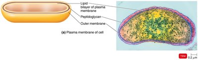
The Cell Wall
Structure and Function
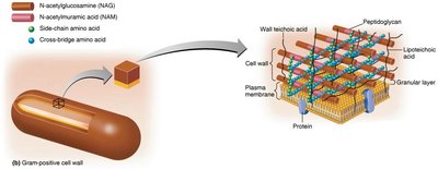
The bacterial cell wall is essential for maintaining cell shape, preventing osmotic lysis, and contributing to pathogenicity. Its composition varies between Gram-positive and Gram-negative bacteria.
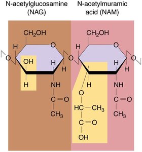
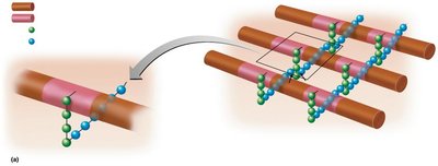
Peptidoglycan: A polymer of N-acetylglucosamine (NAG) and N-acetylmuramic acid (NAM) linked by polypeptides.
Gram-Positive Cell Walls:
Thick peptidoglycan layer
Teichoic acids (wall and lipoteichoic acids) provide rigidity and antigenic specificity
2 rings in basal body of flagella
High susceptibility to penicillin; disrupted by lysozyme
Gram-Negative Cell Walls:
Thin peptidoglycan layer
Outer membrane contains lipopolysaccharide (LPS), lipoproteins, and phospholipids
Lipid A (endotoxin), O polysaccharide (antigenic)
4 rings in basal body of flagella
Low susceptibility to penicillin
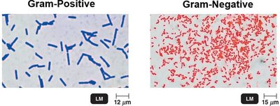
Gram Stain Mechanism
The Gram stain differentiates bacteria based on cell wall structure:
Gram-Positive: Retain crystal violet-iodine complex; appear purple.
Gram-Negative: Lose crystal violet-iodine complex after alcohol wash; counterstained with safranin and appear pink/red.
Atypical Cell Walls
Acid-Fast Cell Walls: Contain mycolic acid; found in Mycobacterium and Nocardia.
Mycoplasmas: Lack cell walls; have sterols in plasma membrane.
Archaea: May lack cell walls or have walls of pseudomurein.
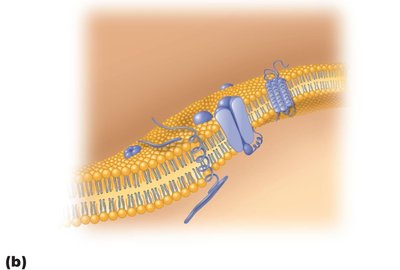
The Plasma (Cytoplasmic) Membrane
Structure and Function
The plasma membrane is a phospholipid bilayer with embedded proteins, following the fluid mosaic model. It is selectively permeable and involved in ATP production and, in some prokaryotes, photosynthesis.
Damage: Alcohols, detergents, and antibiotics can disrupt the membrane, causing leakage of cell contents.
Movement of Materials Across Membranes
Passive Processes
Simple Diffusion: Movement from high to low concentration until equilibrium is reached.
Facilitated Diffusion: Transport of ions and larger molecules via transporter proteins.
Osmosis: Movement of water across a selectively permeable membrane.
Osmotic Pressure and Solutions
Isotonic: Equal solute concentration inside and outside; no net water movement.
Hypotonic: Lower solute outside; water enters cell, may cause lysis.
Hypertonic: Higher solute outside; water leaves cell, causing plasmolysis.
Active Processes
Active Transport: Moves substances against concentration gradient using ATP and transporter proteins.
Group Translocation: Substance is chemically modified during transport (unique to prokaryotes).
The Cytoplasm in Prokaryotes
Internal Structures
Cytoplasm: Mostly water with proteins, carbohydrates, lipids, and ions.
Nucleoid: Contains the bacterial chromosome (circular DNA).
Plasmids: Small, extrachromosomal DNA molecules; often carry antibiotic resistance genes.
Ribosomes: Sites of protein synthesis; 70S (50S + 30S subunits).
Inclusions: Reserve deposits (e.g., metachromatic granules, polysaccharide granules, lipid inclusions, sulfur granules, carboxysomes, gas vacuoles, magnetosomes).
Endospores: Highly resistant, dormant structures formed by Bacillus and Clostridium during nutrient depletion.
Structures Only Found in Eukaryotic Cells
Membrane-Bound Organelles
Nucleus: Contains DNA complexed with histones; site of transcription.
Endoplasmic Reticulum (ER): Rough ER (with ribosomes) synthesizes proteins; smooth ER synthesizes lipids and stores calcium.
Golgi Apparatus: Modifies, sorts, and packages proteins and lipids for secretion or delivery to other organelles.
Lysosomes: Contain digestive enzymes.
Peroxisomes: Break down fatty acids and detoxify harmful substances.
Mitochondria: Site of ATP production via cellular respiration; contain their own DNA and 70S ribosomes.
Chloroplasts: Site of photosynthesis in plants and algae; contain thylakoids and their own DNA.
Centrosomes and Centrioles: Organize microtubules during cell division (animal cells).
Vacuoles: Storage and structural support (especially in plant cells).
Other Eukaryotic Structures
Flagella and Cilia: Composed of microtubules in a 9+2 arrangement; flagella are longer and fewer, cilia are shorter and numerous.
Cell Wall: Present in plants (cellulose), fungi (chitin), and some protists; absent in animal cells.
Glycocalyx: Carbohydrate-rich layer for protection and cell recognition (animal cells).
Plasma Membrane: Contains sterols and carbohydrates; capable of endocytosis (phagocytosis and pinocytosis).
Cytoskeleton: Provides structural support and facilitates movement.
The Evolution of Eukaryotic Structures
Endosymbiotic Theory
The endosymbiotic theory proposes that eukaryotic organelles such as mitochondria and chloroplasts originated from free-living prokaryotes engulfed by ancestral eukaryotic cells. Evidence includes the presence of 70S ribosomes and circular DNA in these organelles.
Additional info: This summary integrates and expands upon the provided lecture notes, including definitions, examples, and academic context for clarity and completeness.
