 Back
BackGram Staining: Principles, Procedure, and Interpretation
Study Guide - Smart Notes
 Tailored notes based on your materials, expanded with key definitions, examples, and context.
Gram Staining
Principle of Differential Staining
The Gram stain is a differential staining technique that distinguishes bacteria based on the structural differences in their cell walls. This method utilizes at least three chemical reagents applied sequentially to a heat-fixed bacterial smear. The outcome of the stain is critical for bacterial classification and identification in microbiology.
Primary Stain (Crystal Violet): Stains all bacterial cells purple.
Mordant (Gram’s Iodine): Forms an insoluble complex with the crystal violet, enhancing retention in certain cell types.
Decolorizing Agent (95% Ethyl Alcohol): Acts as a solvent and protein dehydrating agent. Its effect depends on cell wall composition, cell age, and exposure time. It is the most critical step, as it determines whether the primary stain is retained or removed.
Counterstain (Safranin): Stains decolorized cells pinkish-red, providing contrast to the purple-stained cells.
Key Point: Gram-positive bacteria retain the crystal violet-iodine complex and appear purple, while Gram-negative bacteria lose the primary stain during decolorization and take up the safranin, appearing pink/red.
Gram Staining Procedure
The following steps outline the standard Gram staining protocol used in microbiology laboratories:
Prepare a mixed smear on a clean, labeled slide using a drop of distilled water and small amounts of two different organisms at designated positions.
Allow the smear to air dry or use a slide warmer, then heat-fix the smear.
Flood the smear with Gram’s crystal violet for 1 minute.
Rinse with distilled water.
Apply Gram’s iodine for 1 minute.
Rinse with distilled water.
Decolorize with 95% ethyl alcohol for 15–20 seconds (maximum).
Rinse with distilled water.
Counterstain with safranin for 2 minutes.
Rinse with distilled water.
Blot dry with bibulous paper.
Examine the slide under oil immersion microscopy.
Note: Proper timing during the decolorization step is essential to avoid false results.
Interpretation of Gram Stain Results
Gram staining differentiates bacteria into two major groups based on cell wall structure:
Gram-positive bacteria: Thick peptidoglycan layer retains the crystal violet-iodine complex, appearing purple under the microscope (e.g., Bacillus cereus, Staphylococcus aureus).
Gram-negative bacteria: Thin peptidoglycan layer and outer membrane allow the crystal violet-iodine complex to be washed out, so cells take up the safranin and appear pink/red (e.g., Escherichia coli).
Example: In a mixed smear, Escherichia coli (Gram-negative bacilli) will appear pink, while Bacillus cereus (Gram-positive bacilli) will appear purple.
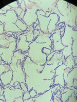
Microscopic Appearance of Gram Stained Bacteria
The image below shows a typical Gram stain result, where Gram-positive bacteria appear purple due to retention of the crystal violet-iodine complex. The morphology and arrangement of cells can also be observed, aiding in identification.
Summary Table: Gram Stain Reagents and Their Functions
Step | Reagent | Function |
|---|---|---|
1 | Crystal Violet | Primary stain; colors all cells purple |
2 | Gram's Iodine | Mordant; forms complex with crystal violet |
3 | 95% Ethyl Alcohol | Decolorizer; removes stain from Gram-negative cells |
4 | Safranin | Counterstain; stains decolorized cells pink/red |
Additional info:
The Gram stain is fundamental for bacterial classification and guides initial clinical treatment decisions.
Over-decolorization can cause Gram-positive cells to appear Gram-negative, while under-decolorization can cause Gram-negative cells to appear Gram-positive.
