 Back
BackHost Defenses and Innate Immunity: An Overview
Study Guide - Smart Notes
 Tailored notes based on your materials, expanded with key definitions, examples, and context.
Host Defenses: Overview
Three Lines of Host Defense
The human body employs a multilevel network of defenses to protect against microbial invasion. These defenses are categorized into three lines: the first and second lines are innate (nonspecific), while the third line is adaptive (specific).
First Line of Defense: Physical and chemical barriers that prevent pathogen entry at portals of entry. These are general and nonspecific.
Second Line of Defense: Internalized system involving protective cells and fluids, including inflammation and phagocytosis. Acts rapidly after the first line is breached.
Third Line of Defense: Adaptive immunity acquired through exposure to specific pathogens, involving lymphocytes and the production of unique protective substances. Provides long-term immunity.
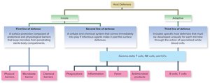
Additional info: The three lines of defense work together, often overlapping and providing redundancy to maximize protection.
Immunology and the Immune System
Definition and Mandate
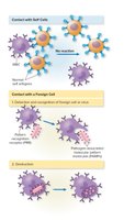
Immunology is the study of the body's second and third lines of defense, including responses to infectious agents, allergies, and cancer. The immune system is responsible for:
Surveillance of the body
Recognition of foreign material
Destruction of entities deemed foreign
Markers and Antigens
Cells are evaluated by the immune system through markers (also called antigens), which are molecules on cell surfaces composed of proteins and/or sugars. These markers help immune cells distinguish between self and nonself, a process central to immune function. Errors in this process can lead to autoimmune disorders.
PAMPs and PRRs
Pathogen-Associated Molecular Patterns (PAMPs): Common molecular signatures found on many microbes but not on mammalian cells. Examples include peptidoglycan and lipopolysaccharide (bacteria), and double-stranded RNA (viruses).
Pattern Recognition Receptors (PRRs): Host cell receptors (on phagocytes, dendritic cells, etc.) that recognize PAMPs and trigger innate immune responses.
Body Systems in Immunity
Major Participating Systems
Mononuclear Phagocyte System (MPS): Network of connective tissue fibers and phagocytic cells in direct contact with tissue cells and extracellular fluid.
Blood: Contains plasma, serum, and formed elements (red blood cells, white blood cells, platelets).
Lymphatic System: Closely connected with the circulatory system, facilitating the movement of immune cells and molecules throughout the body.
Blood Cells in Immunity
Blood cells originate from stem cells in the bone marrow through hematopoiesis. White blood cells (leukocytes) are divided into granulocytes and agranulocytes, both essential for nonspecific and specific immunity.
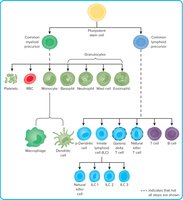
Key Blood Cells in Innate Immunity
Neutrophils: General-purpose phagocytes, first responders to infection, and primary component of pus.
Monocytes: Differentiate into macrophages and dendritic cells in tissues.
Macrophages: Engulf and destroy pathogens, present antigens, and regulate immune responses.
Cytokines: Cell Communication Molecules
Cytokines are small, active molecules secreted by various cells (monocytes, macrophages, lymphocytes, etc.) to regulate, stimulate, or suppress immune responses, inflammation, and cell development. Examples include pro-inflammatory cytokines (e.g., interleukin-1) and anti-inflammatory cytokines.
First Line of Defense: Physical and Chemical Barriers
Skin and Mucous Membranes
The first line of defense consists of inborn, nonspecific barriers that impede the entry of microbes. These include:
Skin: The stratum corneum is a tough, waterproof outer layer. Shedding of skin and hair, as well as sweat, helps remove microbes.
Mucous Membranes: Found in the digestive, urinary, respiratory tracts, and eyes. Mucus impedes entry and attachment of microbes, while blinking, tears, saliva, vomiting, and defecation help flush out pathogens.
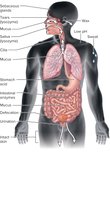
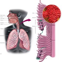
Respiratory Tract Defenses
Nasal hair traps particles.
Mucus and ciliated epithelium move trapped particles toward the pharynx.
Sneezing and coughing expel irritants.
Genitourinary Tract Defenses
Continuous urine flow and periodic bladder emptying flush the urethra.
Vaginal secretions cleanse the lower reproductive tract.
Human Microbiome
The normal microbiota forms a structural barrier, competes with pathogens for nutrients, and alters local pH to inhibit pathogen growth. Disruption of the microbiome can contribute to diseases such as Crohn’s disease and ulcerative colitis.
Second Line of Defense: Cellular and Chemical Systems
Phagocytosis
Phagocytosis is a cornerstone of inflammation and specific immunity. Phagocytes survey tissues, ingest and eliminate microbes, and process antigens. Main phagocytes include neutrophils, monocytes, and macrophages.
Histiocytes: Tissue-resident macrophages (e.g., alveolar macrophages in the lungs, Kupffer cells in the liver).
Steps of Phagocytosis
Chemotaxis: Movement toward chemical signals.
Ingestion: Engulfment of the microbe.
Phagolysosome Formation: Fusion of phagosome with lysosome.
Destruction: Enzymatic and chemical killing of the microbe.
Excretion: Removal of debris.
Recognition of Microbes
PAMPs: Signal molecules on microbes recognized as 'red flags' by phagocytes.
PRRs: Host receptors that bind PAMPs and trigger immune responses.
Inflammasomes: Cytoplasmic PRRs in phagocytes that regulate inflammation upon PAMP recognition.
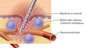
Inflammatory Response
Signs and Symptoms
Rubor: Redness
Calor: Warmth
Tumor: Swelling
Dolor: Pain
Loss of function
Inflammation can be local or systemic, acute or chronic. It is triggered by infection, tissue injury, or immune reactions.
Functions of Inflammation
Mobilize and attract immune components to injury site
Repair tissue damage and localize harmful substances
Destroy microbes and block further invasion
Stages of Inflammation
Vasoconstriction and release of chemical mediators
Vasodilation, increased permeability, and migration of white blood cells
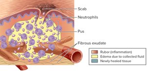
Formation of pus and tissue repair
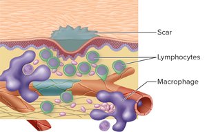
Resolution and scar formation
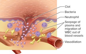
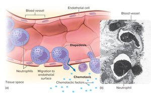
Diapedesis and Chemotaxis
Diapedesis: Migration of white blood cells out of blood vessels into tissues.
Chemotaxis: Movement of cells toward chemical signals at infection sites.
Benefits of Edema and Leaky Vessels
Dilution of toxins
Fibrin clots trap microbes
Neutrophils destroy bacteria and debris
Pus formation (pyogenic bacteria attract neutrophils)
Fever
Definition and Types
Fever is an abnormally elevated body temperature, commonly associated with infection. The hypothalamus maintains body temperature around 37°C (98.6°F).
Low-grade fever: 37.7–38.3°C (100–101°F)
High-grade fever: 40.0–41.4°C (104–106°F)
Pyrogens
Exogenous pyrogens: Derived from outside the body (e.g., microbial products, vaccines).
Endogenous pyrogens: Released by immune cells during phagocytosis (e.g., interleukin-1, tumor necrosis factor).
Benefits and Treatment of Fever
Inhibits growth of temperature-sensitive microbes
Reduces iron availability for bacteria
Increases metabolism and immune activity
Treatment is recommended for high, prolonged fevers or in vulnerable individuals
Antimicrobial Products
Interferons
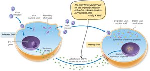
Interferons (IFNs) are small proteins produced by white blood and tissue cells. They regulate immune responses and have antiviral and antitumor effects.
IFN-α and IFN-β: Produced by lymphocytes, fibroblasts, and macrophages; stimulate phagocytes.
IFN-γ: Produced by T cells; regulates macrophages and lymphocytes.
Interferons bind to cell surfaces, induce antiviral proteins, degrade viral RNA, and block viral protein synthesis. They are not microbe-specific and are used therapeutically for some viral infections.
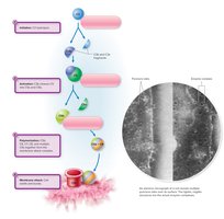
Complement System
The complement system consists of over 30 blood proteins that work in a cascade to destroy bacteria, viruses, and infected cells. The cascade involves:
Initiation: C3 hydrolysis
Activation and cascade: C3b cleaves C5
Polymerization: Formation of membrane attack complex (MAC)
Membrane attack: MAC forms pores, leading to cell lysis
There are two main pathways:
Classical pathway: Initiated by antibodies bound to microbes.
Alternative pathway: Initiated by foreign antigens, faster response.
Antimicrobial Peptides
Short proteins (12–50 amino acids) such as defensins can insert into bacterial membranes, forming pores that lead to cell lysis. They also modulate immune responses and are being explored as therapeutic agents.
