 Back
BackInnate Immunity: Mechanisms and Cellular Components
Study Guide - Smart Notes
 Tailored notes based on your materials, expanded with key definitions, examples, and context.
Innate Immunity
Overview of Immunity
Immunity refers to the body's ability to resist and eliminate pathogens. It is divided into two main types: innate immunity (non-specific, present from birth) and adaptive immunity (specific, acquired after exposure). Innate immunity provides the first line of defense against any pathogen, while adaptive immunity targets specific invaders and provides long-term protection.
Susceptibility: Lack of resistance to a disease.
Immunity: Ability to ward off disease.
Innate Immunity: Defenses against any pathogen, present from birth.
Adaptive Immunity: Immunity or resistance to a specific pathogen, acquired through infection or vaccination.
First Line of Defense: Barriers
The first line of innate immunity consists of physical and chemical barriers that prevent pathogen entry.
Physical Barriers:
Skin: Epidermis of tightly packed cells with keratin, a protective protein.
Mucus: Traps microbes; ciliary escalator moves trapped microbes away from the lungs.
Saliva and Urine: Wash microbes off surfaces and out of the body.
Chemical Barriers:
Fatty acids in sebum: Fungistatic properties.
Low pH: Skin (3–5), gastric juice (1.2–3.0).
Lysozyme: Enzyme in perspiration, tears, saliva, and urine; especially effective against Gram-positive bacteria.
Second Line of Defense: Internal Defenses
If pathogens bypass the first line, the second line involves cellular and protein-based responses.
Cells: Natural killer (NK) cells, phagocytes (macrophages, neutrophils).
Proteins: Complement proteins, interferons.
Response: Recognize general features of pathogens, attack, and destroy them.
Leukocytes (White Blood Cells)
Types and Functions
Leukocytes are the main cellular defenders against infection and foreign invaders. They are classified as granulocytes or agranulocytes based on the presence of granules in their cytoplasm.
Granulocytes:
Neutrophils: Perform phagocytosis; first responders to bacterial infections (60–70% of WBCs).
Basophils: Release histamine; involved in inflammation and allergic reactions (0.5–1%).
Eosinophils: Kill parasites and modulate allergic responses (2–4%).
Agranulocytes:
Lymphocytes: B cells produce antibodies; T cells destroy infected/cancerous cells and coordinate immune responses (20–25%).
Monocytes: Become macrophages in tissues; phagocytose bacteria, dead cells, and debris (3–8%).
Other Blood Components: Thrombocytes (platelets) are responsible for clotting; erythrocytes (red blood cells) transport oxygen and carbon dioxide.
Phagocytosis
Mechanism of Phagocytosis
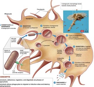
Phagocytosis is the process by which phagocytes (e.g., macrophages, neutrophils) ingest and destroy microbes. The process involves chemotaxis, adherence, ingestion, and digestion.
Phagocyte: The cell that performs phagocytosis.
Phagosome: Vesicle formed around the ingested microbe.
Lysosome: Organelle containing digestive enzymes that fuses with the phagosome to form a phagolysosome.
Key Steps:
Chemotaxis and adherence of microbe to phagocyte.
Ingestion of microbe by phagocyte (formation of phagosome).
Fusion of phagosome with lysosome to form phagolysosome.
Digestion of ingested microbe by enzymes.
Discharge of waste materials.
Microbial Evasion of Phagocytosis
Some pathogens have evolved mechanisms to evade destruction by phagocytes:
Inhibit Adherence: M proteins or capsules prevent phagocyte attachment (e.g., Streptococcus pyogenes, Streptococcus pneumoniae).
Kill Phagocytes: Production of leukocidins that destroy phagocytes (e.g., Staphylococcus aureus).
Lyse Phagocytes: Trigger formation of membrane attack complex to rupture phagocyte membrane (e.g., Listeria monocytogenes).
Escape from Phagosome: Bacteria escape into cytoplasm before destruction (e.g., Shigella, Rickettsia).
Prevent Phagosome/Lysosome Fusion: Block fusion, avoiding digestion (e.g., HIV, Mycobacterium tuberculosis).
Survive Inside Phagolysosome: Resist digestion and survive in harsh conditions (e.g., Coxiella burnetii).
Inflammation Process
Mechanisms and Mediators
Inflammation is a complex response to infection or injury, characterized by redness, swelling, pain, and heat. It is mediated by various proteins and chemicals that increase blood flow and recruit immune cells to the site of infection.
Acute-phase proteins: Produced by the liver in response to inflammation (e.g., histamine, kinins, prostaglandins, leukotrienes, cytokines).
Vasodilation: Widening of blood vessels, increasing blood flow and causing redness (erythema).
Increased permeability: Allows fluid, proteins, and immune cells to enter tissues, causing swelling (edema).
Pain: Caused by swelling and chemical mediators stimulating nerve endings.
Heat: Local increase due to more blood flow; fever is a systemic response mediated by cytokines.
Complement System
Pathways of Complement Activation
The complement system is a group of proteins that enhance immune responses by promoting inflammation, opsonization, and direct lysis of pathogens. There are three main activation pathways:
Pathway | Trigger | Purpose |
|---|---|---|
Classical | Antibodies bound to pathogen | Opsonization, MAC formation, inflammation |
Lectin | Mannose-binding lectin binds to sugars on pathogen | Opsonization, MAC formation, inflammation |
Alternative | Direct activation by pathogen surfaces | Opsonization, MAC formation, inflammation |
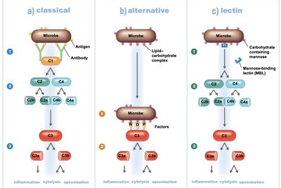
Key Outcomes: Inflammation, cytolysis (cell lysis), and opsonization (enhanced phagocytosis).
How Some Bacteria Evade Complement
Capsules: Prevent activation of the complement system.
Surface lipid-carbohydrates: Block membrane attack complex (MAC) formation.
Discourage opsonization: Seen in Salmonella, Neisseria gonorrhoeae, Bordetella pertussis, Haemophilus influenzae.
Enzymatic digestion of C5a: Some Gram-positive bacteria degrade complement proteins.
Interferons
Types and Functions
Interferons (IFNs) are signaling proteins released by cells in response to infection, especially viral infections.
IFN-α and IFN-β (Alpha and Beta Interferons): Released by infected cells to warn neighboring cells and inhibit viral replication.
IFN-γ (Gamma Interferon): Released by T cells and NK cells; activates macrophages and neutrophils to enhance phagocytosis and microbial killing.
Summary Table: Leukocyte Types and Functions
Leukocyte Type | Main Function | Relative Abundance (%) |
|---|---|---|
Neutrophils | Phagocytosis, first responders | 60–70 |
Lymphocytes | Antibody production, cell-mediated immunity | 20–25 |
Monocytes | Become macrophages, phagocytosis | 3–8 |
Eosinophils | Kill parasites, modulate allergy | 2–4 |
Basophils | Release histamine, allergy, inflammation | 0.5–1 |
Key Equations and Concepts
Opsonization: The process by which pathogens are marked for ingestion and destruction by phagocytes.
Membrane Attack Complex (MAC): A structure formed on the surface of pathogen membranes that leads to cell lysis.
Phagocytosis Equation (Simplified):
Complement Activation (Classical Pathway):
