 Back
BackInnate Immunity: The Body’s First and Second Lines of Defense
Study Guide - Smart Notes
 Tailored notes based on your materials, expanded with key definitions, examples, and context.
Innate Immunity
Overview of Host Defenses
Innate immunity provides the body’s immediate, nonspecific defense against pathogens. It consists of physical barriers, cellular responses, and chemical mediators that act as the first and second lines of defense to prevent and control infections.
The Body’s First Line of Defense
Physical and Chemical Barriers
Skin and mucous membranes are the primary structures preventing pathogen entry.
These barriers are supported by antimicrobial chemicals and processes.
The Role of Skin in Innate Immunity
Skin is composed of two major layers:
Epidermis: Multiple layers of tightly packed cells; few pathogens can penetrate. Shedding of dead cells removes microorganisms. Epidermal dendritic cells phagocytize pathogens.
Dermis: Contains collagen fibers that resist abrasions, preventing microbial entry.
Chemical defenses of skin:
Perspiration: Contains salt (inhibits microbial growth), antimicrobial peptides, and lysozyme (destroys bacterial cell walls).
Sebum: Secreted by sebaceous glands; keeps skin pliable and lowers pH, inhibiting many bacteria.
The Role of Mucous Membranes in Innate Immunity
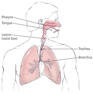
Mucous membranes line all body cavities open to the environment (respiratory, digestive, urinary, reproductive tracts).
Consist of two layers:
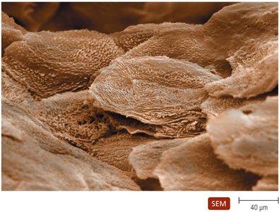
Epithelium: Thin, living, tightly packed cells; continual shedding removes microbes. Dendritic cells below epithelium phagocytize pathogens. Goblet and ciliated columnar cells help remove invaders.
Deeper connective tissue: Supports the epithelium and produces antimicrobial chemicals.
The Role of the Lacrimal Apparatus
Lacrimal apparatus produces and drains tears, washing the eye surface.
Tears contain lysozyme, which destroys bacteria.
The Role of the Microbiome
Microbial antagonism: The normal microbiome competes with potential pathogens by:
Consuming nutrients
Creating unfavorable environments for pathogens
Preventing pathogen attachment to host cells
Stimulating the body’s second line of defense
Producing antimicrobial compounds
Providing vitamins and modulating immunity
Other First-Line Defenses
Antimicrobial peptides: Present in skin, mucous membranes, and neutrophils; act against a variety of microbes by disrupting membranes or interfering with metabolism.
Other organs secrete chemicals with antimicrobial properties.
The Body’s Second Line of Defense
General Features
The second line of defense is activated when pathogens penetrate the skin or mucous membranes. It includes cellular components, antimicrobial chemicals, and physiological processes, many of which are found in the blood.
Defense Components of Blood
Plasma: Mostly water, containing electrolytes, dissolved gases, nutrients, proteins (including iron-binding compounds, complement proteins, and antibodies).
Serum: Plasma without clotting factors.
Formed elements:
Erythrocytes: Transport oxygen and carbon dioxide.
Platelets: Involved in blood clotting.
Leukocytes: Defend against invaders; divided into granulocytes and agranulocytes.
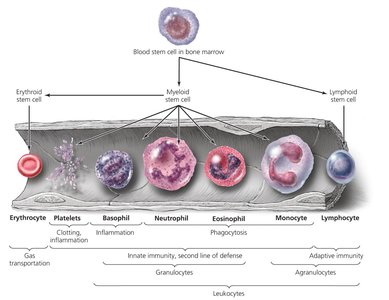
Leukocytes: Granulocytes
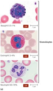
Contain large granules that stain differently:
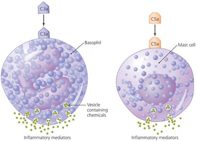
Basophils: Stain blue; release inflammatory chemicals.
Eosinophils: Stain red/orange; phagocytize pathogens and attack helminths.
Neutrophils: Stain lilac; phagocytize pathogens and can kill without phagocytosis.
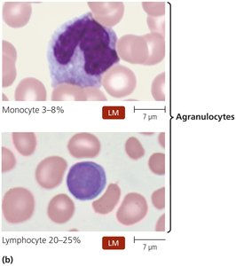
Leukocytes: Agranulocytes
Cytoplasm appears uniform under light microscopy:
Lymphocytes: Most involved in adaptive immunity; includes natural killer (NK) cells.
Monocytes: Leave blood and mature into macrophages, which are phagocytic.
Lab Analysis of Leukocytes
Differential white blood cell counts can indicate disease:
Increased eosinophils: Allergies or parasitic worm infection
Increased leukocytes/neutrophils: Bacterial infection
Increased lymphocytes: Viral infection
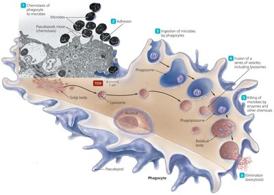
Phagocytosis
Phagocytosis is the process by which certain cells (phagocytes) ingest and destroy pathogens. It occurs in six stages:
Chemotaxis
Adhesion
Ingestion
Maturation
Killing
Elimination
Nonphagocytic Killing
Eosinophils: Attack helminths by secreting toxins; can form extracellular structures that kill bacteria.
Natural Killer (NK) cells: Secrete toxins onto virally infected cells and tumors; distinguish normal cells by surface proteins.
Neutrophils: Produce chemicals and extracellular traps (NETs) to kill microbes without phagocytosis.
Nonspecific Chemical Defenses Against Pathogens
Toll-like receptors (TLRs): Membrane proteins on phagocytes that recognize pathogen-associated molecular patterns (PAMPs) and trigger defensive responses (e.g., inflammation, apoptosis).
NOD proteins: Cytosolic proteins that bind PAMPs and trigger inflammation and apoptosis.
Interferons: Proteins released by host cells to inhibit viral spread; cause symptoms of viral infections. Two types: Type I (alpha, beta) and Type II (gamma).
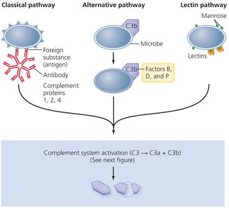
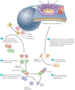
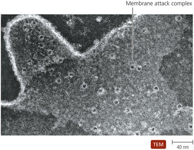
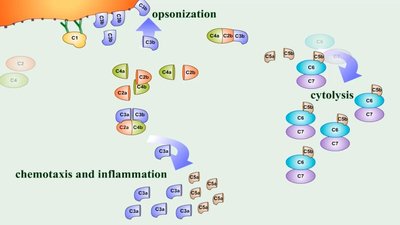
Complement system: A set of serum proteins that, when activated, result in lysis of foreign cells, inflammation, and opsonization. Activation occurs via classical, alternative, or lectin pathways.
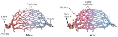
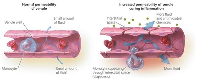
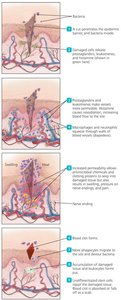
Inflammation
Inflammation is a nonspecific response to tissue damage, characterized by redness, heat, swelling, and pain. It can be acute (short-lived, beneficial) or chronic (long-lasting, potentially damaging).
Acute inflammation: Increases blood flow and vascular permeability, recruits phagocytes, and promotes tissue repair.
Chronic inflammation: Can cause tissue damage and disease.
Chemicals involved: Bradykinins, prostaglandins, leukotrienes, histamine.
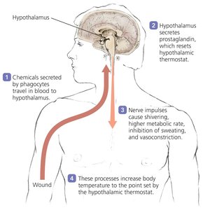
Fever
Fever is a body temperature above 37°C, triggered by pyrogens that reset the hypothalamic thermostat. Pyrogens include bacterial toxins, cytoplasmic contents of lysed bacteria, antibody-antigen complexes, and pyrogens released by phagocytes.
Fever enhances interferon effects, inhibits some microbes, and may enhance phagocyte and immune cell activity and tissue repair.
Summary Table: Nonspecific Components of Innate Immunity
First Line | Second Line |
|---|---|
Barriers and Associated Chemicals: Skin and mucous membranes prevent entry of pathogens; chemicals (e.g., sweat, acid, lysozyme, mucus) enhance protection. | Phagocytes: Macrophages, neutrophils, and eosinophils ingest and destroy pathogens. Extracellular Killing: Eosinophils and NK lymphocytes kill pathogens without phagocytosis; neutrophils can also kill without phagocytosis. Complement: Attracts phagocytes, stimulates inflammation, and attacks pathogen membranes. Interferons: Increase resistance to viral infection, slow disease spread. Antimicrobial Peptides: Disrupt membranes, signaling, and metabolism. Inflammation: Increases blood flow, capillary permeability, and leukocyte migration; walls off infection; increases local temperature. Fever: Mobilizes defenses, accelerates repair, inhibits pathogens. |
