 Back
BackChapter One
Study Guide - Smart Notes
 Tailored notes based on your materials, expanded with key definitions, examples, and context.
Chapter 1: The Microbial World
What is Microbiology?
Microbiology is the scientific study of microorganisms, also known as microbes. These organisms are typically microscopic and can be found in nearly every environment on Earth. Microbes include both prokaryotic and eukaryotic organisms, and they play essential roles in various biological and ecological processes.
Microbes are found almost everywhere, from soil and water to extreme environments.
They can be prokaryotes (Bacteria and Archaea) or eukaryotes (such as fungi, protozoa, and algae).
Some microbes are capable of photosynthesis, while others produce antibiotics or have commercial applications.
Microbiology is fundamental to the study of molecular biology, biochemistry, genomics, and molecular genetics.
Diversity and Classification of Microbes
Microbes are highly diverse in both form and function. They inhabit every environment that supports life, ranging from single-celled organisms to complex multicellular structures. Microbes often live in communities and interact with each other and their environment.
Most microbes are harmless or beneficial; only a minority are pathogenic (disease-causing).
Microbes are classified using a binomial nomenclature (genus and species), e.g., Escherichia coli, Bacillus anthracis.
Beneficial Activities of Microbes
While some microbes are known for causing infectious diseases, many are beneficial and essential for life on Earth. Their activities include:
Nutrient cycling (e.g., nitrogen fixation in soil)
Decomposition of organic matter
Fermentation in food production (e.g., cheese, yogurt, bread)
Production of antibiotics and other pharmaceuticals
Bioremediation (cleaning up pollutants)
Industrial applications (e.g., biofuels, enzymes)

Microbes in Agriculture and the Human Body
Modern agriculture relies on microbes for soil fertility and plant health, especially through nitrogen fixation.
The human gastrointestinal tract contains a vast community of microbes essential for digestion and health.


Microbes in Food and Industry
Microbes are crucial in the production of fermented foods and industrial processes.
Fermentation by microbes produces foods such as cheese, yogurt, sauerkraut, and alcoholic beverages.
Industrial microbiology uses microbes to produce chemicals, antibiotics, and biofuels.


Tools for Studying Microbes
Microbiologists use a variety of tools and techniques to study microbes, including:
Microscopy: Essential for visualizing microbes due to their small size.
Culturing: Growing microbes in or on nutrient media to study their properties.
Colony: A visible mass of microbial cells arising from a single cell or group of cells.
Molecular techniques: Such as 16S rRNA gene sequencing for identifying and classifying microbes.

Evolutionary Relationships and the Tree of Life
Microbes are classified into three domains: Bacteria, Archaea, and Eukarya. Phylogenetic relationships are determined using molecular data, especially ribosomal RNA sequences.
The phylogenetic tree of life shows evolutionary relationships among all living organisms.
Molecular techniques allow scientists to analyze genetic material and construct evolutionary trees.

Variation in Microbial Size and Shape
Microbes exhibit a wide range of sizes and shapes, which can affect their physiology and ecological roles.
Microbial cells can be spherical, rod-shaped, spiral, or filamentous.
Size impacts surface area-to-volume ratio, influencing nutrient uptake and growth rates.


Properties of Microbial Cells
All microbial cells share certain fundamental properties, while some have specialized features.
Structure: All cells have a cytoplasmic membrane, cytoplasm, ribosomes, and DNA.
Metabolism: Cells use genetic information to produce proteins and carry out biochemical reactions.
Growth: Cells increase in number through division.
Evolution: Genetic changes allow adaptation and speciation.
Differentiation: Some cells form specialized structures (e.g., spores).
Communication: Cells interact via chemical signals.
Motility: Some cells can move using flagella or other structures.
Horizontal gene transfer: Exchange of genetic material between cells.

Microbial Contribution to Global Biomass
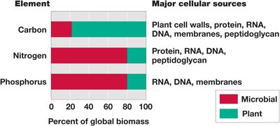
Microbes play a significant role in the Earth's biomass and biogeochemical cycles, contributing to the cycling of carbon, nitrogen, and phosphorus.
Element | Major Cellular Sources | Percent of Global Biomass (Microbial) | Percent of Global Biomass (Plant) |
|---|---|---|---|
Carbon | Plant cell walls, protein, RNA, DNA, membranes, peptidoglycan | ~20% | ~80% |
Nitrogen | Protein, RNA, DNA, peptidoglycan | ~80% | ~20% |
Phosphorus | RNA, DNA, membranes | ~80% | ~20% |

History of Microbiology: Early Microscopy
The field of microbiology began with the invention of the microscope. Early pioneers include:
Robert Hooke (1635–1703): First to describe microbes and illustrated mold structures.
Antoni van Leeuwenhoek (1632–1723): First to observe bacteria using a simple microscope.

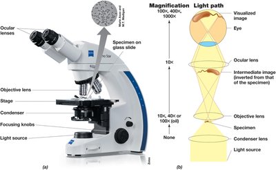
Light Microscopy and Staining
Light microscopy is a fundamental tool for observing microbes. Staining techniques improve contrast, making cells and their structures more visible.
Bright-field microscopy: Standard method for viewing stained cells.
Staining: Involves fixing and coloring cells to enhance visibility.

Advanced Light Microscopy Techniques
Several advanced microscopy techniques are used to improve contrast and visualize live or unstained cells:
Phase-contrast microscopy: Enhances contrast in unstained, live cells by exploiting differences in refractive index.
Fluorescence microscopy: Uses fluorescent dyes or natural fluorescence to visualize specific cell components; widely used in diagnostics and ecology.
Differential interference contrast (DIC) microscopy: Uses polarized light to give a three-dimensional appearance to cells.
Dark-field microscopy: Produces bright images on a dark background, useful for observing motility.

Electron Microscopy
Electron microscopy (EM) uses electrons instead of light to achieve much higher resolution, allowing visualization of cellular ultrastructure.
Transmission Electron Microscopy (TEM): Electrons pass through thin specimens, revealing internal structures at the molecular level.
Scanning Electron Microscopy (SEM): Electrons are scattered from the surface, providing detailed 3D images of cell surfaces and large specimens.
Negative staining allows direct observation of intact cells and components.
Robert Koch and the Germ Theory of Disease
Robert Koch (1843–1910) established the link between microbes and infectious diseases, laying the foundation for medical microbiology.
Identified causative agents of anthrax, tuberculosis, and cholera.
Formulated Koch’s postulates to definitively link specific microbes to specific diseases.
Developed solid media for obtaining pure cultures and observed that colonies with different characteristics bred true.
Koch’s Postulates:
The suspected pathogen must be present in all cases of the disease and absent from healthy organisms.
The suspected pathogen must be isolated and grown in pure culture.
Cells from a pure culture of the suspected pathogen must cause disease in a healthy organism.
The pathogen must be re-isolated from the newly diseased organism and shown to be the same as the original.
Additional info: Koch’s work established the scientific method for identifying the microbial causes of infectious diseases, which remains foundational in microbiology and medicine.
