Back
BackLymphatic System and Immunity: Structure, Function, and Clinical Importance
Study Guide - Smart Notes
 Tailored notes based on your materials, expanded with key definitions, examples, and context.
Lymphatic System and Immunity
Introduction to the Immune System
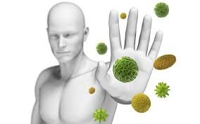
The immune system is a complex network that protects the body from toxins, bacteria, viruses, and cancerous cells. It acts as a safety net, shielding us from disease-causing agents through specialized cells and molecules that provide effective and specific resistance to disease.
Key Functions: Maintains fluid balance and defends against infection and disease.
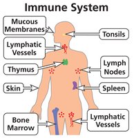
Components: Lymphatic organs (fixed structures), defensive cells, and molecules (mobile protectors).
The Lymphatic System
Organization and Role of the Lymphatic System
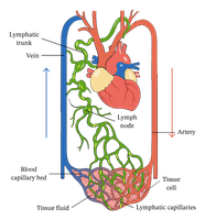
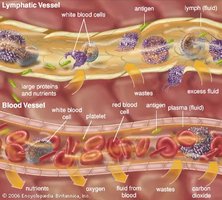
The lymphatic system maintains fluid balance around body cells and works closely with the cardiovascular system. Blood delivers nutrients and removes waste in capillary beds, but some fluid and proteins remain in tissues after capillary exchange. This excess becomes lymph, which is returned to the bloodstream via lymphatic vessels.
Lymph: Tissue fluid, proteins, lipids, and other substances.
Function: Recovers lost fluid and prevents edema (swelling).
Structure & Components of the Lymphatic System
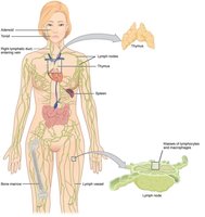
The lymphatic system consists of lymph, lymphatic vessels, lymph nodes, and lymphoid organs (thymus and spleen). These components act as biological filters, removing harmful particles and preventing potential damage to tissues and organs.
Lymphatic Vessels: Transport lymph and filter it through lymph nodes.
Lymph Nodes: Filter lymph before it returns to the bloodstream.
Lymphoid Organs: Thymus and spleen, which develop immune cells and provide defense against infections.
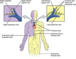
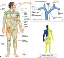
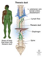
Major Lymphatic Ducts
The lymphatic system includes two main ducts:
Right lymphatic duct: Drains the upper right body and empties into the right subclavian vein.
Thoracic duct: Drains the rest of the body and empties into the left subclavian vein.
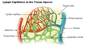
Formation and Role of Lymph
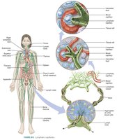
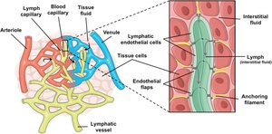
Lymph forms from blood plasma that filters out of blood capillaries into tissue spaces due to hydrostatic pressure. This fluid mixes with substances from tissue cells, creating interstitial fluid (IF). Excess IF drains into lymphatic capillaries, where it is called lymph. Without drainage, excess IF would cause swelling (edema).
Key Point: Lymphatic capillaries are tiny, blind-ended tubes found throughout tissue spaces.
Contents: Lymph may contain proteins, waste, lipids, and other substances.


Role of the Lymphatic System in Fluid Balance
Lymphatic vessels transport lymph, filtering it through lymph nodes. This process prevents fluid buildup, helps maintain fluid homeostasis, and returns clean, filtered lymph to the bloodstream.
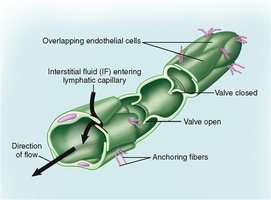
Lymphatic Vessels: Structure and One-Way Flow
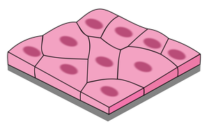
Lymphatic capillaries are microscopic vessels made of simple squamous epithelium (endothelium) with looser cell junctions than blood capillaries, allowing larger molecules to enter. Lymph flows in one direction only, aided by valves that prevent backflow and help lymph move toward the heart.
Flow Path: Lymphatic capillaries → lymphatic venules → lymphatic veins → lymphatic ducts → bloodstream.
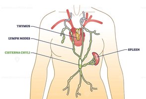
The Two Main Lymphatic Ducts
The thoracic duct drains three-quarters of the body and empties into the left subclavian vein, beginning in the abdomen at the cisterna chyli. The right lymphatic duct drains the right upper body and empties into the right subclavian vein. Both ducts return filtered lymph to the bloodstream.
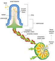
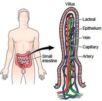
Special Role of Lacteals
Lacteals are special lymphatic capillaries in the small intestine that absorb lipids (fats) from digested food and transport them into the bloodstream, playing a vital role in nutrient absorption.
Lymphoid Organs
Overview of Lymphoid Organs
Lymphoid organs include lymph nodes, thymus, tonsils, and spleen. They contain lymphoid tissue made up of developing lymphocytes and other immune-related cells supported by reticular fibers. These organs are essential for developing immune cells and providing defense against infections.
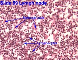
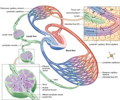
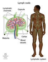
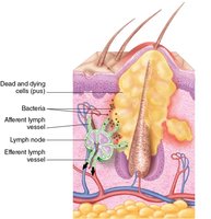
Lymph Nodes: Structure and Function
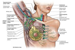
Lymph nodes are small, bean-shaped structures found along lymphatic vessels. They filter lymph before it returns to the bloodstream and are usually found in clusters in the neck, armpits, groin, chest, and abdomen.
Structure: Capsule (outer covering), cortex (outer area), medulla (inner area), reticular fibers (mesh), afferent vessels (bring lymph in), efferent vessels (carry lymph out).
Filtration: Mechanical filtration traps debris; biological filtration uses immune cells (phagocytes) to destroy bacteria, viruses, and cancer cells.
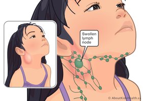
Clinical Importance of Lymph Nodes
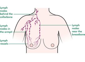
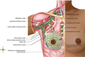
Swollen lymph nodes are a sign of infection. Breast lymph drainage is clinically important, as it can help drain milk and also spread cancer or infection. Axillary lymph nodes are often removed in breast cancer surgery to prevent metastasis.
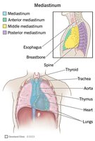
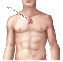
The Thymus: Structure and Function
The thymus is located in the mediastinum and extends toward the neck. It is largest at puberty and shrinks with age. The thymus is made of lymphocytes and reticular tissue and is responsible for T cell maturation and hormone secretion (thymosins).
T Cell Maturation: Immature lymphocytes from fetal bone marrow finish developing in the thymus.
Hormone Secretion: Thymosins help T cell development.
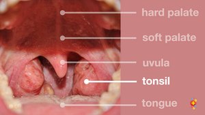
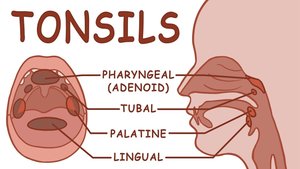
Tonsils: Structure, Location, and Types
Tonsils are masses of lymphoid tissue located in a protective ring under mucous membranes of the mouth and throat. They are positioned around the openings of the nasal and oral cavities and form a defensive ring in the pharyngeal region.
Types: Palatine tonsils (each side of the throat), pharyngeal tonsils (adenoids, near posterior nasal cavity), lingual tonsils (base of the tongue).
Main Functions: Immune surveillance and first line of defense against airborne or ingested microbes.

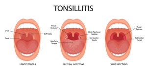
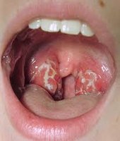
Clinical Importance of Tonsils
Tonsillitis is a common infection due to high exposure; chronic or obstructive tonsillitis may require removal (tonsillectomy). Even after removal, the immune system adapts as other immune tissues help protect the body.
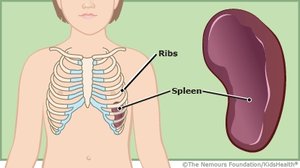
The Spleen: Structure and Function
The spleen is located in the upper left abdomen, lateral to the stomach, and protected by lower ribs. It contains a large network of veins and can hold over 500 mL of blood.
Main Functions: Filtration (removes bacteria and debris from blood), RBC recycling (destroys old RBCs and salvages iron), blood reservoir (stores blood for emergency use).
Immune Role: Contains white pulp with lymphocytes, stores monocytes for rapid response, supports tissue repair during injury or infection.
Clinical Importance: Splenectomy (surgical removal) may be necessary after trauma or rupture. The body can function without the spleen, but infection risk increases, requiring vaccinations and preventive care.