 Back
BackChapter Two
Study Guide - Smart Notes
 Tailored notes based on your materials, expanded with key definitions, examples, and context.
Microbial Cell Structure and Function
Overview of the Prokaryotic Cell Envelope
The cell envelope of prokaryotes is a complex, multi-layered structure that surrounds the cytoplasm and mediates interactions with the environment. It is essential for maintaining cell integrity, shape, and protection against external stresses.
Cytoplasmic membrane: The innermost boundary, crucial for selective permeability and metabolic functions.
Cell wall: Provides rigidity and protection from osmotic lysis.
Outer membrane: Present in Gram-negative bacteria, adds an extra layer of protection.
S-layers: Paracrystalline protein or glycoprotein layers, often the outermost structure in some Archaea and Bacteria.
Cytoplasmic Membrane Structure and Function
Bacterial Cytoplasmic Membrane
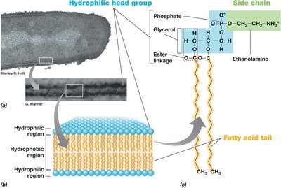
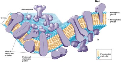
The bacterial cytoplasmic membrane is a selectively permeable phospholipid bilayer with embedded proteins. It separates the cytoplasm from the external environment and is fundamental for cellular homeostasis.
Composed of phospholipids with hydrophilic heads and hydrophobic tails.
Integral and peripheral proteins are involved in transport, energy generation, and signal transduction.
Archaeal Cytoplasmic Membrane
The archaeal cytoplasmic membrane differs significantly from that of bacteria, reflecting adaptations to extreme environments.
Contains ether linkages (instead of ester) between glycerol and hydrophobic side chains.
Hydrophobic side chains are isoprenes rather than fatty acids.
Major lipids include phosphoglycerol diethers with phytanyl side chains and diphosphoglycerol tetraethers with biphytanyl side chains, which can form monolayers for increased stability.
Functions of the Cytoplasmic Membrane
The cytoplasmic membrane is essential for several cellular processes:
Permeability barrier: Prevents leakage and controls the passage of nutrients and waste.
Protein anchor: Site for proteins involved in transport, bioenergetics, and chemotaxis.
Energy conservation: Site of generation and dissipation of the proton motive force.

Transport Across the Membrane
Transport systems are vital for nutrient uptake and waste removal. They include:
Simple diffusion: Movement of small, nonpolar molecules down their concentration gradient.
Facilitated diffusion and active transport: Use of membrane proteins to transport substances, often against gradients.
High-affinity transporters: Saturate at lower substrate concentrations, allowing efficient uptake in nutrient-poor environments.


The Cell Wall
General Functions
The cell wall is a rigid structure that protects the cell from osmotic lysis, maintains shape, and is a key determinant in bacterial classification (Gram stain).
Composed mainly of peptidoglycan in bacteria.
Provides mechanical strength and defines cell morphology.
Peptidoglycan Structure
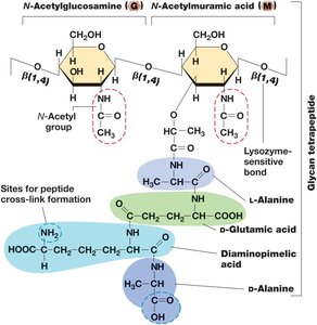
Peptidoglycan is a unique macromolecule forming a mesh-like layer outside the cytoplasmic membrane in most bacteria.
Consists of alternating N-acetylglucosamine (G) and N-acetylmuramic acid (M) residues linked by β(1,4) glycosidic bonds.
Peptide cross-links provide additional strength.

Gram-Positive vs. Gram-Negative Cell Walls
Bacteria are classified based on cell wall structure:
Gram-positive: Thick peptidoglycan layer, teichoic and lipoteichoic acids, susceptible to lysozyme and penicillin.
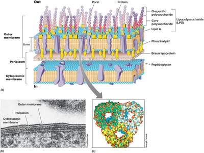
Gram-negative: Thin peptidoglycan layer, outer membrane with lipopolysaccharide (LPS), periplasmic space.


Archaeal Cell Walls
Archaeal cell walls differ fundamentally from those of bacteria:
Lack peptidoglycan; some contain pseudomurein (with β(1,3) linkages).
Many have an S-layer as the main cell wall component.

S-Layers
S-layers are crystalline protein or glycoprotein layers that provide structural support and protection.
Always the outermost layer if present.
Functions include protection from lysis, maintaining shape, and mediating cell interactions.

Surface Structures: Capsules, Slime Layers, Pili, and Fimbriae
Capsules and Slime Layers
These are polysaccharide-rich layers external to the cell wall, important for surface attachment and protection.
Capsule: Tightly attached, well-organized matrix.
Slime layer: Loosely attached, easily deformed.
Functions: Biofilm formation, virulence, prevention of desiccation.

Pili and Fimbriae
These are thin, proteinaceous appendages that facilitate attachment, motility, and genetic exchange.
Fimbriae: Short, numerous, used for attachment to surfaces.
Pili: Longer, fewer, involved in conjugation (genetic exchange) and motility (e.g., type IV pili).
Some pili are electrically conductive (nanowires).

Hami (Archaeal Appendages)
Hami are unique, hook-like appendages found in some Archaea, facilitating attachment in extreme environments.

Cell Inclusions
Types and Functions
Cell inclusions are intracellular storage granules that serve as energy reserves or reservoirs for specific elements.
Enclosed by a thin protein membrane.
Reduce osmotic stress and store carbon, phosphorus, sulfur, or other minerals.
Carbon Storage Polymers
Common carbon storage polymers include:
Poly-β-hydroxyalkanoate (PHA): Accumulates when carbon is in excess, used as a carbon and energy source.
Glycogen: Glucose polymer, also serves as a carbon and energy reservoir.

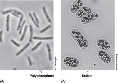
Polyphosphate, Sulfur, and Carbonate Minerals
Other inclusions include:
Polyphosphate granules: Store inorganic phosphate.
Elemental sulfur granules: Store sulfur, which can be oxidized to sulfate.
Carbonate minerals: Biomineralization of barium, strontium, and magnesium.

Gas Vesicles
Gas vesicles are protein-bound structures that confer buoyancy, allowing aquatic bacteria to position themselves optimally in the water column.
Composed of two proteins forming conical, gas-filled structures.

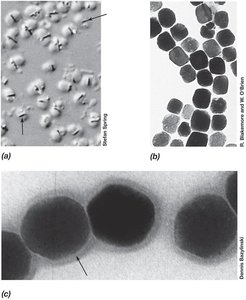
Magnetosomes
Magnetosomes are intracellular, membrane-bound crystals of magnetic iron minerals that enable bacteria to orient along magnetic field lines (magnetotaxis).
Allow navigation in aquatic environments.

Endospores
Structure and Function
Endospores are highly differentiated, dormant cells formed by some Gram-positive bacteria (e.g., Bacillus and Clostridium) to survive extreme conditions.
Resistant to heat, radiation, chemicals, and desiccation.
Serve as survival structures during unfavorable growth conditions.
Ideal for dispersal via wind, water, or animal gut.
Endospore Formation and Germination
Endospore formation (sporulation) is a complex, multistage process involving asymmetric cell division, engulfment, cortex and coat formation, and maturation.
Germination returns the endospore to a vegetative state when conditions improve.

Differences Between Endospores and Vegetative Cells
The following table summarizes key differences between vegetative cells and endospores:
Characteristic | Vegetative Cell | Endospore |
|---|---|---|
Microscopic appearance | Nonrefractile | Refractile |
Calcium content | Low | High |
Dipicolinic acid | Absent | Present |
Enzymatic activity | High | Low |
Respiration rate | High | Low or absent |
Macromolecular synthesis | Present | Absent |
Heat resistance | Low | High |
Radiation resistance | Low | High |
Resistance to chemicals | Low | High |
Lysozyme | Sensitive | Resistant |
Water content | High, 80–90% | Low, 10–25% in core |
Small acid-soluble spore proteins | Absent | Present |
