 Back
BackMicrobial Diseases of the Digestive System: Structure, Pathogenesis, and Clinical Features
Study Guide - Smart Notes
 Tailored notes based on your materials, expanded with key definitions, examples, and context.
Microbial Diseases of the Digestive System
Overview of the Digestive System
The digestive system is a complex series of organs and glands that processes food, extracts nutrients, and eliminates waste. It is a major site for microbial colonization and infection due to its exposure to ingested materials.
Main organs: Oral cavity, pharynx, esophagus, stomach, small intestine, large intestine, rectum, anus
Accessory organs: Salivary glands, liver, gallbladder, pancreas
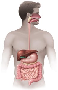
Normal microbiota are abundant, especially in the mouth and large intestine, and play roles in digestion and immune defense.
Normal Microbiota and Host Defenses
Normal Microbiota
Millions of bacteria per milliliter of saliva
Large numbers in the large intestine (up to 100 billion bacteria per gram of feces)
Host Defenses
Stomach: Acidic environment inhibits many pathogens
Small intestine: Paneth cells produce α-defensins and other antimicrobial peptides
Bacterial Diseases of the Mouth
Structure of the Tooth
Teeth are susceptible to microbial colonization due to their non-shedding surfaces, allowing biofilm (dental plaque) to accumulate.
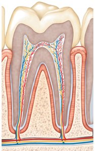
Enamel: Hard, outermost layer
Dentin: Underlies enamel, less mineralized
Pulp cavity: Contains nerves and blood vessels
Gingiva: Gum tissue surrounding teeth
Dental Caries (Tooth Decay)
Streptococcus mutans is the primary pathogen responsible for dental caries. It metabolizes carbohydrates, tolerates acidity, and synthesizes dextrans that help form biofilms.
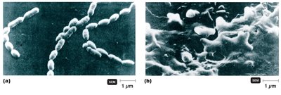
Biofilm formation: S. mutans adheres to tooth surfaces, especially in the presence of sucrose, producing sticky dextran polymers.
Acid production: Fermentation of sugars produces acids that demineralize enamel.
Stages of Tooth Decay
Tooth decay progresses through several stages, beginning with plaque formation and advancing to pulp involvement if untreated.
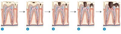
Healthy tooth with plaque
Decay in enamel
Advanced decay
Decay in dentin
Decay in pulp
Periodontal Disease
Periodontal diseases affect the supporting structures of the teeth, including the gums and bone. Porphyromonas species are major pathogens.
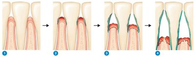
Gingivitis: Inflammation of the gums
Periodontitis: Destruction of supporting bone and periodontal ligament, formation of periodontal pockets
Summary Table: Bacterial Diseases of the Mouth
Disease | Pathogen |
|---|---|
Dental caries | Streptococcus mutans |
Periodontal disease | Porphyromonas spp. |
Acute necrotizing gingivitis | Prevotella intermedia |
Bacterial Diseases of the Lower Digestive System
General Concepts
Infection: Growth of a pathogen in the GI tract (incubation: 12 hours to 2 weeks)
Intoxication: Ingestion of preformed toxins (symptoms: 1 to 48 hours after ingestion)
Gastroenteritis: Inflammation of the stomach and intestines, causing diarrhea and dysentery
Treatment: Oral rehydration therapy is often essential
Staphylococcal Food Poisoning
Pathogen: Staphylococcus aureus
Symptoms: Nausea, vomiting, diarrhea
Mechanism: Intoxication by enterotoxin (superantigen)
Diagnosis: Phage typing
Treatment: None (self-limiting)
Shigellosis (Bacillary Dysentery)
Pathogen: Shigella spp.
Symptoms: Tissue damage, dysentery
Mechanism: Infection; produces endotoxin and Shiga exotoxin
Diagnosis: Isolation of bacteria
Treatment: Fluoroquinolones
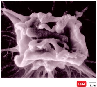
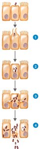
Salmonellosis and Typhoid Fever
Salmonellosis Pathogen: Salmonella enterica
Typhoid Fever Pathogen: Salmonella typhi
Symptoms: Nausea, diarrhea (salmonellosis); high fever, significant mortality (typhoid)
Mechanism: Infection; endotoxin production
Diagnosis: Isolation and serotyping
Treatment: Oral rehydration (salmonellosis); quinolones/cephalosporins (typhoid)
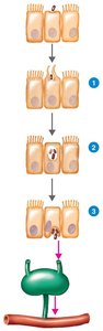
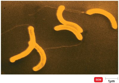
Cholera and Other Vibrios
Cholera Pathogen: Vibrio cholerae O:1 and O:139
Symptoms: Profuse watery diarrhea ("rice-water stools")
Mechanism: Cholera toxin (exotoxin) causes secretion of Cl-, HCO3-, and water
Treatment: Rehydration, doxycycline
Escherichia coli Gastroenteritis
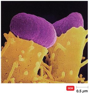
EPEC: Enteropathogenic E. coli – forms pedestals on host cells
EIEC: Enteroinvasive E. coli – invades intestinal mucosa
EAEC: Enteroaggregative E. coli – produces enterotoxin, watery diarrhea
EHEC: Enterohemorrhagic E. coli – produces Shiga toxin, can cause hemolytic uremic syndrome (HUS)
Helicobacter Peptic Ulcer Disease
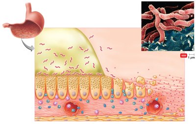
Pathogen: Helicobacter pylori
Symptoms: Peptic ulcers
Mechanism: Infection; urease production neutralizes stomach acid
Diagnosis: Urea breath test, bacterial culture
Treatment: Antimicrobial drugs
Viral Diseases of the Digestive System
Mumps
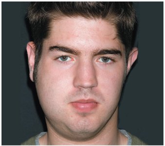
Pathogen: Mumps virus
Symptoms: Swollen parotid glands
Incubation: 16–18 days
Treatment: Preventive vaccine
Viral Hepatitis
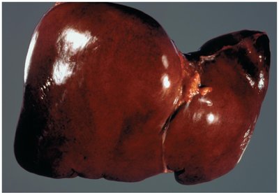
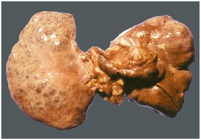
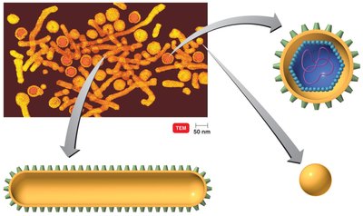
Hepatitis is inflammation of the liver, caused by several viruses (A, B, C, D, E) with different transmission routes and outcomes.
Disease | Transmission | Pathogen | Chronic Liver Disease? | Vaccine? |
|---|---|---|---|---|
Hepatitis A | Fecal-oral | Picornaviridae | No | Inactivated virus |
Hepatitis B | Parenteral, STI | Hepadnaviridae | Yes | Recombinant |
Hepatitis C | Parenteral | Filoviridae | Yes | None |
Hepatitis D | Parenteral, HBV coinfection | Deltaviridae | Yes | HBV vaccine |
Hepatitis E | Fecal-oral | Caliciviridae | No | HAV vaccine |
Fungal Diseases of the Digestive System
Mycotoxin Intoxications
Disease | Pathogen | Symptoms | Intoxication/Infection | Diagnosis | Treatment |
|---|---|---|---|---|---|
Ergot Poisoning | Claviceps purpurea | Reduced blood to limbs | Mycotoxin in grain | Sclerotia in food | None |
Aflatoxin Poisoning | Aspergillus flavus | Liver cirrhosis; liver cancer | Mycotoxin in food | Immunoassay for toxin in food | None |
Protozoan Diseases of the Digestive System
Giardiasis
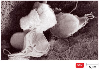
Pathogen: Giardia lamblia
Symptoms: Diarrhea due to protozoan adherence to intestinal wall
Reservoir: Water or mammals
Diagnosis: FA test
Treatment: Metronidazole; quinacrine
Cryptosporidiosis
Pathogen: Cryptosporidium hominis
Symptoms: Self-limiting diarrhea; severe in immunosuppressed
Reservoir: Cattle; water
Diagnosis: Acid-fast stain; FA; ELISA
Treatment: Oral rehydration
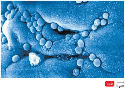
Amebic Dysentery
Pathogen: Entamoeba histolytica
Symptoms: Abscesses, significant mortality
Reservoir: Humans
Diagnosis: Microscopy; serology
Treatment: Metronidazole
Helminthic Diseases of the Digestive System
Overview of Helminths
Kingdom: Animalia
Phyla: Platyhelminthes (flatworms: trematodes/flukes, cestodes/tapeworms), Nematoda (roundworms)
Features: Chemoheterotrophic, multicellular, complex life cycles, reduced digestive/nervous systems
Life Cycle of Helminths
Monoecious: Both male and female reproductive organs in one animal
Dioecious: Separate male and female individuals
Stages: Egg → larva(e) → adult
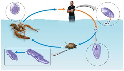
Clonorchis sinensis (Liver Fluke)
Transmission: Ingestion of undercooked, infested fish
Symptoms: Often asymptomatic; fever, abdominal pain, jaundice
Diagnosis: Eggs in fecal matter
Treatment: Praziquantel
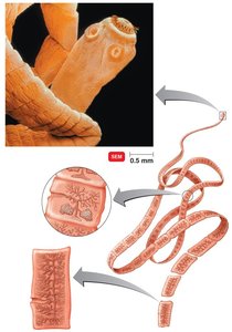
Taenia spp. (Tapeworms)
T. saginata: Beef tapeworm
T. solium: Pork tapeworm
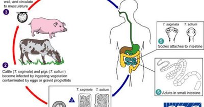
Transmission: Ingestion of undercooked meat containing larvae
Definitive host: Humans
Intermediate host: Cattle (T. saginata), pigs (T. solium)
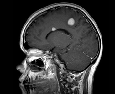
Neurocysticercosis
Neurocysticercosis is a severe complication of T. solium infection, where larvae invade the central nervous system, causing neurological symptoms.
Additional info: For all diseases, prevention includes proper food handling, sanitation, and, where available, vaccination. Oral rehydration is a cornerstone of treatment for diarrheal diseases.
