 Back
BackMicrobiology Foundations: History, Cell Types, Microscopy, and Lab Techniques
Study Guide - Smart Notes
 Tailored notes based on your materials, expanded with key definitions, examples, and context.
Introduction to Microbiology
Historical Timeline and Development of Microbiology
Microbiology developed as a science through key discoveries and technological advances. The timeline below highlights major contributors and their findings:
1600s – Robert Hooke: First to publish descriptions of cells using a microscope.
1632–1723 – Antonie van Leeuwenhoek: First to observe bacteria and protozoa, calling them “animalcules.”
1840s – Ignaz Semmelweis: Introduced handwashing in hospitals to prevent infections.
1860s – Joseph Lister: Developed antiseptic surgery using carbolic acid.
1822–1895 – Louis Pasteur: Supported biogenesis, invented pasteurization, and developed vaccines for rabies and anthrax.
1843–1910 – Robert Koch: Established germ theory, formulated Koch’s postulates, and identified disease-causing microbes.
1850–1920 – Golden Age of Microbiology: Major discoveries in culturing and identifying pathogens.
Key findings: Microorganisms cause disease, produce food, make medications, break down hazards, and inhabit nearly every environment on Earth.
Theories of Spontaneous Generation and Biogenesis
Spontaneous generation: The belief that life arises from nonliving matter (e.g., maggots from meat).
Biogenesis: Life arises only from existing life.
Evidence against spontaneous generation: Redi’s covered meat jars (no maggots) and Pasteur’s swan-neck flask (no microbial growth unless exposed to air).
Major Contributors to Microbiology
Louis Pasteur: Disproved spontaneous generation, developed pasteurization, and created vaccines.
Robert Koch: Developed germ theory, Koch’s postulates, and improved staining/culturing methods.
Joseph Lister: Introduced antiseptic surgery and sterilization techniques.
Ignaz Semmelweis: Advocated hand hygiene, reducing childbed fever.
Robert Hooke: First to describe cells under a microscope.
Antonie van Leeuwenhoek: First to observe living bacteria and protozoa.
Florence Nightingale: Established aseptic nursing techniques and modern nursing.
Characteristics and Classification of Microorganisms
Features of Living Things and Non-Cellular Microbes
Viruses: Acellular, nonliving, composed of genetic material (DNA or RNA) and a protein coat (capsid); some have a lipid envelope. Require a host cell to replicate.
Distinguishing Features of Microbial Groups
Algae: Photosynthetic protists with chlorophyll, found in aquatic environments.
Bacteria: Unicellular prokaryotes lacking a nucleus.
Fungi: Eukaryotes (yeasts, molds) with chitin cell walls.
Protozoans: Unicellular eukaryotes, classified by movement.
Viruses: Acellular infectious particles.
Helminths: Parasitic worms (roundworms and flatworms).
Classification of Bacteria and Fungi; Mycosis
Bacteria: Classified by shape, size, arrangement, Gram stain, and metabolism.
Fungi: Classified by yeast/mold form, hyphae structure, and spore formation.
Mycosis: Fungal infection or disease.
Bacterial Endospores vs. Fungal Spores
Bacterial endospore: Dormant survival structure (e.g., Bacillus, Clostridium); highly resistant to heat, chemicals, and radiation.
Fungal spore: Reproductive structure for growth and spread.
Healthcare challenge: Endospores are difficult to kill and may persist on medical equipment, increasing infection risk.
Classification of Protozoans and Helminths
Protozoans: Classified by motility (amoeboid, flagellated, ciliated, spore-forming).
Helminths: Two main groups: roundworms (nematodes) and flatworms (platyhelminths); classified by body shape.
Microscopy and Staining Techniques
Types of Microscopes and Techniques
Light microscope: Uses light to view cells and bacteria.
Transmission Electron Microscope (TEM): Shows internal cell structures (2D image).
Scanning Electron Microscope (SEM): Shows cell surface (3D image).
Staining: Enhances visibility of cells and structures.
Oil immersion: Improves image clarity by reducing light refraction.
Key Microscopy Terms
Total magnification: Product of ocular and objective lens magnifications.
Resolution: Ability to distinguish two close objects as separate.
Refractive index: Measure of how much light bends passing through a substance.
Comparison of TEM and SEM
TEM: Reveals internal structures, 2D images.
SEM: Reveals surface details, 3D images.
Staining Techniques
Simple stain: One dye; shows shape, size, arrangement.
Differential stain: Multiple dyes; distinguishes cell types (e.g., Gram stain).
Structural stain: Highlights specific structures (capsules, spores, flagella).
Clinical application: Identifies bacteria and guides treatment.
Gram Stain Procedure and Interpretation
Steps: crystal violet → iodine → acetone-alcohol → safranin.
Gram-positive: Thick peptidoglycan, retains purple.
Gram-negative: Thin peptidoglycan, turns pink.
Errors: Over-decolorizing, thick smears, old cells.
Acid-Fast Stain
Genera: Mycobacterium, Nocardia.
Reason: Mycolic acid in cell walls.
Clinical use: Detects tuberculosis and related infections.
Microbiome and Microbial Interactions
Healthy Microbiome
Normal microbes on skin, mouth, intestines.
Roles: protect against pathogens, aid digestion, maintain health.
Microbe-Host Interactions
Beneficial: Aid digestion, protect from infection.
Harmful: Cause disease.
Neutral: No effect on host.
Symbiotic Relationships
Parasitism: One benefits, other harmed.
Mutualism: Both benefit.
Commensalism: One benefits, other unaffected.
Biofilms and Microbial Applications
Biofilm Formation and Healthcare Implications
Microbes attach to surfaces, multiply, and produce a protective layer.
Healthcare implications: Biofilms are hard to remove, antibiotic-resistant, and common on medical devices.
Beneficial Roles of Microbes
Bioremediation: Cleaning pollutants (oil spills, toxic waste).
Food production: Yogurt, cheese, bread, beer, wine.
Drug production: Antibiotics, insulin, vitamins, enzymes, biofuels.
Aseptic Techniques and Lab Safety
Aseptic Techniques
Prevent contamination and infection (handwashing, gloves, sterilization, disinfecting).
Lab Safety and PPE
PPE: Gloves, lab coat, goggles, closed-toe shoes.
Allowed: Wearing PPE, disinfecting, aseptic technique, labeling cultures.
Not allowed: Eating/drinking, touching face, horseplay, leaving cultures open.
Disposal of Cultures
Petri plates and test tubes: Place in biohazard waste, autoclave before disposal.
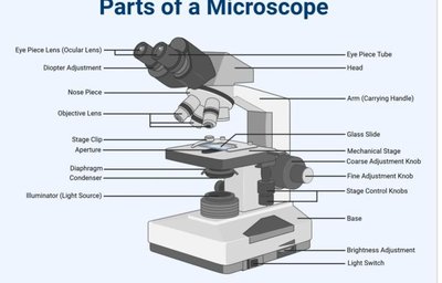
Microscope Structure and Function
Parts of the Compound Light Microscope
Ocular lens (eyepiece): Viewing lens.
Objective lenses: Magnification.
Stage: Holds slide.
Coarse/fine adjustment knobs: Focus.
Light source: Illumination.
Arm/base: Support.
Calculating Total Magnification
Formula:
Example: 10× ocular × 40× objective = 400×
Scientific Method Steps
Ask a question/make observation
Form hypothesis
Perform experiment
Collect data
Analyze results
Draw conclusion
Microbial Growth and Culture Techniques
Best Practices for Aseptic Technique
Wash hands, disinfect workspace, flame loop, keep plates closed, use sterile instruments, wear gloves.
Invert plates: Prevent condensation from spreading bacteria.
Media Types and Formats
General-purpose media: Nutrient agar, nutrient broth.
Formats: Broth, plates, slants, deeps.
Streak Plate Method for Isolation
Sterilize loop
Collect sample
Streak first section
Flame loop
Streak subsequent sections
Invert and incubate
Goal: Isolate individual colonies from single cells.
Biochemistry and Cell Structure
Macromolecules and Chemical Bonds
Dehydration synthesis: Joins molecules by removing water.
Hydrolysis: Breaks molecules by adding water.
Covalent bonds: Share electrons.
Ionic bonds: Transfer electrons.
Hydrogen bonds: Weak attractions between molecules.
Acids, Bases, and pH
Acid: pH < 7
Base: pH > 7
Neutral: pH = 7
Major Macromolecules
Macromolecule | Monomer | Function |
|---|---|---|
Carbohydrates | Monosaccharides | Energy |
Lipids | Fatty acids + Glycerol | Energy, membranes |
Proteins | Amino acids | Enzymes, structure |
Nucleic acids | Nucleotides | Genetic information |
Cell Types and Taxonomy
Bacteria, Archaea, and Eukaryotes
Bacteria: Prokaryotic, peptidoglycan cell wall.
Archaea: Prokaryotic, no peptidoglycan.
Eukaryotes: Nucleus and organelles.
Scientific Naming and Taxonomy
Genus: Capitalized, italicized (e.g., Escherichia).
Species: Lowercase, italicized (e.g., coli).
Hierarchy: Domain → Kingdom → Phylum → Class → Order → Family → Genus → Species
Strain: Variant within a species.
Prokaryotic vs. Eukaryotic Cells
Feature | Prokaryotic | Eukaryotic |
|---|---|---|
Nucleus | No | Yes |
Organelles | No | Yes |
Size | Smaller | Larger |
Binary Fission
DNA replicates → cell grows → septum forms → two identical daughter cells.
Bacterial Structures and Functions
Structure | Function |
|---|---|
Nucleoid | Contains DNA |
Flagella | Movement |
Pili | DNA transfer |
Fimbriae | Attachment |
Ribosome | Protein synthesis |
Capsule | Protection |
Endosymbiotic Theory and Mitochondria
Mitochondria have their own DNA, ribosomes, double membrane, and divide like bacteria, supporting the endosymbiotic theory.
Eukaryotic Cell Structures and Functions
Structure | Function |
|---|---|
Nucleus | Stores DNA |
Rough ER | Protein synthesis |
Smooth ER | Lipid synthesis |
Golgi apparatus | Packages proteins |
Mitochondria | ATP/energy |
Chloroplast | Photosynthesis |
Lysosome | Digestion |
Peroxisome | Detoxification |
Gram-Positive vs. Gram-Negative Cell Walls
Feature | Gram-Positive | Gram-Negative |
|---|---|---|
Peptidoglycan | Thick | Thin |
Color after Gram stain | Purple | Pink |
Outer membrane | No | Yes |
Osmosis and Bacterial Cells
Osmosis: Movement of water across a membrane.
Hypertonic: Water leaves cell, cell shrinks.
Hypotonic: Water enters, cell swells.
Isotonic: No net movement.
Eukaryotic Plasma Membranes
Phospholipid bilayer controls entry/exit.
Animals: Cholesterol
Fungi: Ergosterol
Plants: Phytosterols
