 Back
BackMicrobiology Lab Practical Review: Microscopy, Aseptic Technique, Staining, and Microbial Identification
Study Guide - Smart Notes
 Tailored notes based on your materials, expanded with key definitions, examples, and context.
Microscopy and Observation of Microorganisms
Parts of the Microscope and Magnification
The microscope is an essential tool in microbiology for observing microorganisms that are invisible to the naked eye. Understanding its parts and how to calculate total magnification is fundamental for laboratory work.
Key Parts: Ocular lens (eyepiece), objective lenses, stage, condenser, diaphragm, coarse and fine focus knobs, light source, arm, and base.
Total Magnification: Calculated by multiplying the magnification of the ocular lens by the objective lens used. Formula:
Example: If the ocular lens is 10x and the objective lens is 40x, total magnification is 400x.
Aseptic Technique and Microbial Cultivation
Common Aseptic Transfers and Inoculation Methods
Aseptic technique prevents contamination of cultures and the environment. Proper use of tools and methods is critical for reliable results.
Tools: Inoculating loop, inoculating needle, Bunsen burner, sterile pipettes, and culture media.
Purpose: To transfer microorganisms without introducing contaminants.
Spread Plate and Streak Plate Methods of Isolation
These techniques are used to isolate pure colonies from a mixed culture.
Spread Plate Method: Involves spreading a diluted microbial sample evenly across the surface of an agar plate using a sterile spreader. Purpose: To obtain isolated colonies for enumeration and identification. Materials: Sterile spreader, agar plate, pipette.
Streak Plate Method: Involves streaking a loopful of culture across the agar surface in a pattern that thins out the sample and separates individual cells. Purpose: To obtain isolated colonies. Correct Procedure: Sterilize loop between quadrants, avoid overlapping streaks.
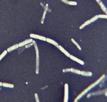
Colony Morphology
Colony morphology describes the visible characteristics of microbial colonies on solid media, which aids in identification.
Key Features: Shape (circular, irregular, filamentous), edge (entire, undulate, lobate), surface (smooth, rough, wrinkled), elevation (flat, raised, convex), color, and size.
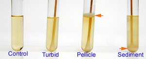
Growth Patterns in Broth
Bacteria exhibit distinct growth patterns in liquid media, which can help in their identification.
Clear: No growth.
Turbid: Cloudy appearance due to dispersed growth.
Flocculent: Clumps of growth suspended in broth.
Pellicle: Growth at the surface.
Sediment: Growth at the bottom.
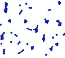
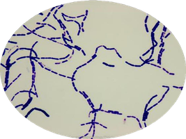
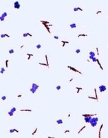
Staining Techniques
Simple Staining
Simple staining uses a single basic dye to highlight cell morphology, size, and arrangement.
Purpose: To visualize bacterial cells and determine their shape and arrangement.
Basic Stains: Crystal violet, methylene blue, safranin.
Charge: Basic stains are positively charged and bind to negatively charged bacterial cell walls.
Heat Fixing: Kills bacteria and adheres them to the slide.
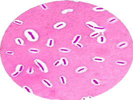
Negative Staining
Negative staining uses acidic dyes to stain the background, leaving cells colorless.
Dyes: Nigrosin, India ink, Eosin, Picric acid.
Charge: Negative (anionic) stains repel the negatively charged cell surface.
Result: Cells appear clear against a dark background.
Gram Staining
Gram staining differentiates bacteria based on cell wall structure into Gram-positive and Gram-negative.
Steps: Primary stain (crystal violet), mordant (iodine), decolorizer (ethyl alcohol), counterstain (safranin).
Gram-positive: Thick peptidoglycan, retain crystal violet (purple).
Gram-negative: Thin peptidoglycan, lose crystal violet, take up safranin (pink/red).
Acid-Fast Staining
Used to identify acid-fast organisms (e.g., Mycobacterium) with waxy cell walls.
Primary Stain: Carbol fuchsin.
Decolorizer: Acid-alcohol.
Counterstain: Methylene blue.
Result: Acid-fast bacteria appear magenta; non-acid-fast appear blue.
Capsule Staining
Capsule staining visualizes the gelatinous capsule surrounding some bacteria.
Capsule Composition: Polysaccharides or polypeptides.
Charge: Neutral.
Result: Capsule appears as a clear halo around stained cells and background.
Endospore Staining
Endospore staining detects bacterial endospores, which are resistant structures.
Primary Stain: Malachite green (stains endospores).
Counterstain: Safranin (stains vegetative cells red).
Result: Endospores appear green, vegetative cells red.
Flagella Staining
Flagella staining visualizes bacterial flagella, which are usually too thin to see with light microscopy.
Types of Flagella: Monotrichous (single), lophotrichous (tuft), amphitrichous (both ends), peritrichous (all over).
Microbial Media and Biochemical Identification
Types of Media
Selective Media: Inhibits growth of some organisms while allowing others (e.g., PEA, MacConkey, EMB, MSA).
Differential Media: Distinguishes organisms based on biochemical reactions (e.g., blood agar, MSA, MacConkey, EMB).
Phenylethyl Alcohol Agar (PEA)
Selective for Gram-positive bacteria; inhibits Gram-negative.
Columbia CNA with Sheep Blood
Selective for Gram-positive; differential for hemolysis (alpha, beta, gamma).
Mannitol Salt Agar (MSA)
Selective for halophiles (e.g., Staphylococcus); differential for mannitol fermentation (yellow = positive).
MacConkey Agar
Selective for Gram-negative; differential for lactose fermentation (pink = positive).
Eosin Methylene Blue (EMB) Agar
Selective for Gram-negative; differential for lactose fermentation (green metallic sheen = E. coli).
Fungi, Protozoa, and Helminths
Common Fungi
Molds: Rhizopus (sporangia, zygospore), Penicillium (conidiospores), Aspergillus, Candida albicans (pseudohyphae).
Yeasts: Saccharomyces cerevisiae (budding yeast).
Protozoa of Clinical Importance
Giardia lamblia (trophozoite), Plasmodium vivax, Entamoeba histolytica, Amoeba, Paramecium.
Parasitic Helminths
Clonorchis sinensis (Chinese liver fluke), Dipylidium caninum (proglottids), Ascaris lumbricoides (adult and egg), Enterobius vermicularis (adult and egg), Taenia solium (scolex and proglottids), Necator americanus (adult and egg), Schistosoma mansoni (egg).
Summary Table: Key Media and Their Properties
Medium | Selective Ingredient | Differential Ingredient | Purpose |
|---|---|---|---|
PEA | Phenylethyl alcohol | None | Gram-positive selection |
Columbia CNA | Colistin, nalidixic acid | Blood/hemoglobin | Gram-positive selection, hemolysis |
MSA | 7.5% NaCl | Mannitol, phenol red | Halophile selection, mannitol fermentation |
MacConkey | Bile salts, crystal violet | Lactose, neutral red | Gram-negative selection, lactose fermentation |
EMB | Eosin Y, methylene blue | Lactose | Gram-negative selection, lactose fermentation |
Additional info:
For all staining methods, always use proper aseptic technique to avoid contamination.
Colony morphology and growth patterns are initial steps in microbial identification, but further biochemical and genetic tests are often required for confirmation.
