 Back
BackMicroscopy in Microbiology: Principles, Types, and Applications
Study Guide - Smart Notes
 Tailored notes based on your materials, expanded with key definitions, examples, and context.
Microscopy: Principles and Parameters
Introduction to Microscopy
Microscopy is a fundamental technique in microbiology, enabling scientists to observe cells and microorganisms that are too small to be seen with the naked eye. The use of microscopes has revolutionized our understanding of microbial structure and function.
Microscope: An instrument that magnifies small objects, allowing detailed visualization.
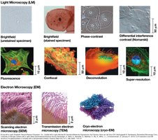
Light Microscope (LM): Uses visible light and glass lenses to magnify specimens.
Electron Microscope (EM): Uses electron beams for much higher resolution.
Key Parameters in Microscopy
Three critical parameters determine the effectiveness of a microscope:
Magnification: The ratio of an object's image size to its real size. Total magnification is calculated as:
Resolution: The ability to distinguish two adjacent points as separate. Higher resolution means clearer images.
Contrast: The difference in brightness or color between a specimen and its background, making structures easier to see.
Refractive Index and Lenses
Refractive Index: Measures how much a substance slows the velocity of light, affecting the direction and magnitude of light bending.
Focal Point: The specific place where light rays are focused by a lens.
Focal Length: Distance between the center of the lens and the focal point; shorter focal length yields higher magnification.
Types of Light Microscopes
Bright-Field Microscope
The bright-field microscope produces a dark image against a bright background and is commonly used for stained specimens.
Multiple objective lenses allow for varying magnification.
Parfocal microscopes remain in focus when objectives are changed.
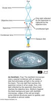
Dark-Field Microscope
The dark-field microscope forms images by light reflected or refracted by the specimen, producing a bright image against a dark background. It is especially useful for observing living, unstained preparations and internal structures in eukaryotic microorganisms.
Used to identify bacteria such as Treponema pallidum.
Phase-Contrast Microscope
Phase-contrast microscopy allows detailed examination of living organisms and internal cell structures without fixation or staining. It brings together direct and diffracted light rays to form an image, enhancing contrast.
Useful for observing cellular structures in their natural state.
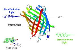
Fluorescence Microscope
Fluorescence microscopy exposes specimens to ultraviolet, violet, or blue light. Specimens are usually stained with fluorochromes, which absorb UV light and emit visible light.
Essential for identifying pathogens and localizing specific proteins in cells.
Fluorochrome-labeled probes (e.g., antibodies) tag cell constituents.
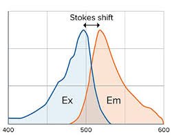
Principle of Fluorescence
Fluorescent substances absorb excitation light and emit light at a longer wavelength (Stokes shift).
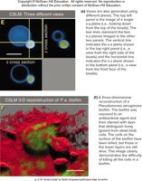
Confocal Microscopy
Confocal microscopy uses fluorochrome dyes and short-wavelength light to excite a single plane of a specimen, producing exceptionally clear two-dimensional images. Multiple planes can be illuminated to construct a three-dimensional image.
Provides high-resolution, 3D reconstructions of cells and tissues.

Electron Microscopy
Transmission Electron Microscope (TEM)
TEM uses electrons that scatter when passing through thin sections of a specimen. Denser regions scatter more electrons and appear darker.
Allows visualization of internal structures at very high resolution.
Scanning Electron Microscope (SEM)
SEM uses electrons excited from the surface of a specimen to create detailed, realistic 3D images of surface features.
Useful for studying microbial morphology and surface structures.
Staining Techniques in Microscopy
Preparing Smears and Fixation
Staining: Coloring microorganisms with dyes to emphasize certain structures.
Smear: Thin film of material containing microorganisms spread over a slide.
Fixation: Attaches and kills microorganisms, preserving their structure.
Heat fixation: Used for bacteria and archaea; preserves morphology.
Chemical fixation: Used for larger, delicate organisms; preserves fine structure.
Types of Stains
Basic Dyes: Positively charged; bind to negatively charged cell components.
Acid Dyes: Negatively charged; bind to positively charged cell components.
Simple Stain: Uses a single dye to determine size, shape, and arrangement.
Differential Stain: Divides microorganisms into groups based on staining properties (e.g., Gram stain, acid-fast stain).
Gram Staining
Most widely used differential staining procedure.
Divides bacteria into Gram-positive and Gram-negative based on cell wall structure.
Acid-Fast Staining
Useful for staining Mycobacterium species (e.g., M. tuberculosis, M. leprae).
High lipid content in cell walls (mycolic acid) is responsible for staining characteristics.
Staining Specific Structures
Capsule Stain: Visualizes polysaccharide capsules; capsules may appear colorless against a stained background.
Flagella Stain: Mordant increases thickness of flagella for visualization.
TEM Specimen Preparation
Specimens must be cut very thin.
Chemically fixed and stained with electron-dense materials (e.g., heavy metals).
Summary Table: Types of Microscopy
Microscope Type | Principle | Application |
|---|---|---|
Bright-field | Light passes through specimen | Stained specimens, general observation |
Dark-field | Light reflected/refracted by specimen | Living, unstained specimens |
Phase-contrast | Direct and diffracted light rays | Internal cell structures, living cells |
Fluorescence | UV light excites fluorochromes | Pathogen identification, protein localization |
Confocal | Laser excites single plane | 3D cell/tissue imaging |
TEM | Electrons pass through thin sections | Internal structure, high resolution |
SEM | Electrons excited from surface | Surface morphology, 3D images |
Additional info: The notes expand on brief points with academic context, definitions, and examples to ensure completeness and clarity for microbiology students.
