 Back
BackMicroscopy, Staining, and Classification in Microbiology
Study Guide - Smart Notes
 Tailored notes based on your materials, expanded with key definitions, examples, and context.
Microscopy, Staining, and Classification
Introduction
This section covers essential techniques in microbiology for visualizing, differentiating, and classifying microorganisms. These methods are foundational for identifying pathogens, understanding microbial structure, and performing laboratory diagnostics.
Urinary Tract Infections (UTIs) and Causative Agents
Overview of UTI Pathogens
Urinary tract infections (UTIs) are among the most common bacterial infections, with most cases being endogenous (originating from the patient's own flora).
Approximately 70% of UTIs are caused by uropathogenic Escherichia coli (E. coli).
Other intestinal bacteria such as Proteus, Klebsiella, and Enterobacter account for about 10% of cases.
Non-intestinal bacteria, including Staphylococcus saprophyticus and Pseudomonas aeruginosa, are responsible for 5–20% of UTIs.
Staining Techniques in Microbiology
Why Stain Microbes?
Most microbes are difficult to see with bright-field microscopy due to their lack of contrast.
Staining increases contrast, making cells and their structures visible.
Preparation involves making a smear, fixing cells, and then applying stains.
Simple Stains
Simple stains use a single basic dye to color cells, allowing visualization of cell size, shape, and arrangement.
Common dyes: crystal violet, safranin, methylene blue.
Method: Fix smear, soak in dye, rinse with water.

Differential Stains
Differential stains use more than one dye to distinguish between different types of cells or cellular components.
Common differential stains include:
Gram stain
Acid-fast stain
Endospore stain
Histological stains
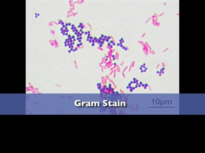
Gram Stain
The Gram stain is the most widely used differential stain in microbiology, separating bacteria into Gram-positive (purple) and Gram-negative (pink) groups based on cell wall structure.
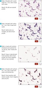
Steps:
Make a smear and fix cells.
Stain with crystal violet, rinse with water.
Add iodine (mordant), rinse with water.
Decolorize with ethanol, rinse with water.
Counterstain with safranin, rinse and dry.
Gram-positive bacteria retain crystal violet and appear purple; Gram-negative bacteria lose crystal violet and take up safranin, appearing pink.
Acid-Fast Stain
The acid-fast stain identifies bacteria with waxy cell walls, such as Mycobacterium and Nocardia. Acid-fast bacteria appear red, while non-acid-fast bacteria are blue.
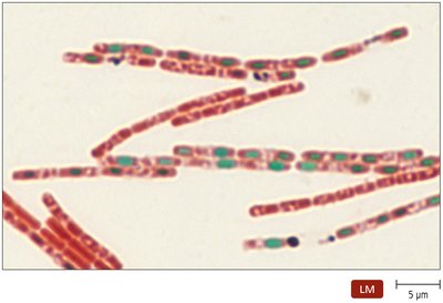
Endospore Stain
This stain is used to detect endospores in genera such as Bacillus and Clostridium. Endospores stain green, while vegetative cells stain red.
Histological Stains
Used for tissue specimens.
Common stains: Gomori methenamine silver (GMS), Hematoxylin and eosin (HE).
Special Stains
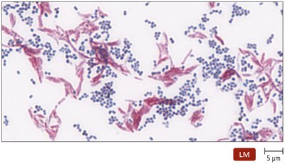
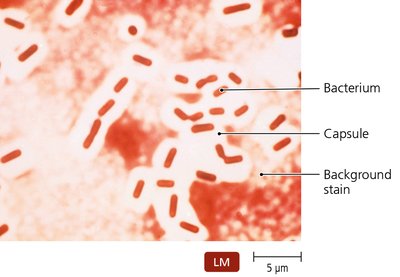
Negative (Capsule) Stain
Negative stains reveal the presence of bacterial capsules, which appear as clear halos around cells against a dark background.
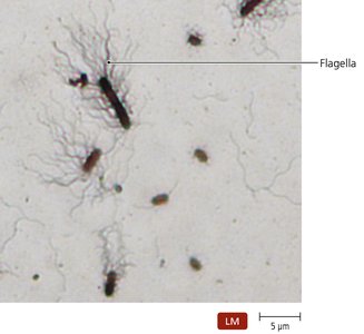
Flagellar Stain
Flagellar stains bind to bacterial flagella, increasing their diameter and making them visible under the microscope. This allows identification of the number and arrangement of flagella.
Taxonomic and Identifying Characteristics
Methods for Identifying Microorganisms
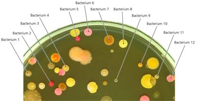
Physical characteristics: Colony morphology, cell shape, and arrangement.
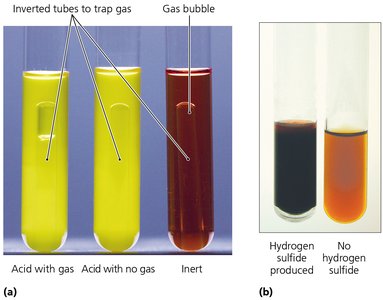
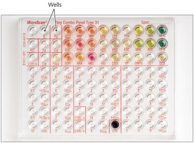
Biochemical tests: Assess metabolic capabilities (e.g., fermentation, enzyme production).
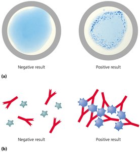
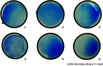
Serological tests: Use antibodies to detect specific microbial antigens.
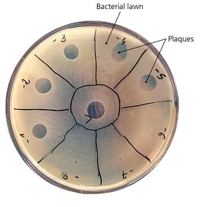
Phage typing: Uses bacteriophages to identify bacterial strains.
MALDI/TOF Mass Spectrometry: Identifies microbes based on protein profiles.
Nucleic acid analysis: Detects and identifies microbes using DNA/RNA sequences.
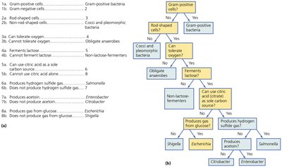
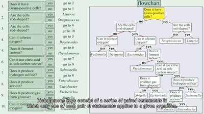
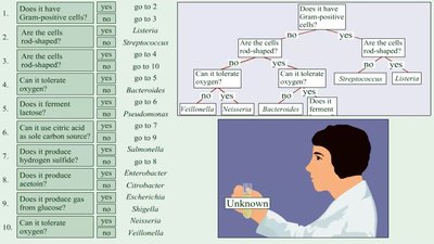
Dichotomous Keys
Using Dichotomous Keys for Microbial Identification
Dichotomous keys are tools that use a series of paired statements to guide users toward the identification of an organism. Each choice leads to another pair of statements or to the identification of the organism.
Review Questions
Serological testing allows lab technicians to test pathogenic bacterial specimens for reaction with known antibodies.
Simple stain is the process that uses a single dye to color a microscope specimen.
Gram stain is the most frequently used differential stain in modern microbiology labs, differentiating microbes into two distinct groups.
