 Back
BackNegative Staining: Principles, Procedure, and Morphology of Micrococcus luteus and Bacillus cereus
Study Guide - Smart Notes
 Tailored notes based on your materials, expanded with key definitions, examples, and context.
Negative Staining in Microbiology
Principles of Negative Staining
Negative staining is a microbiological technique used to visualize cell morphology, size, and arrangement without subjecting cells to distortion from heat fixation. This method employs an acidic stain, such as Nigrosin, which is negatively charged. Because bacterial cell surfaces are also negatively charged, the stain is repelled and does not penetrate the cells. As a result, cells appear transparent against a dark background, allowing for accurate observation of their natural characteristics.
Acidic stains (e.g., Nigrosin) are repelled by the negatively charged bacterial cell surface.
Cells remain unstained and are visible as clear areas against a black background.
No heat fixation is used, preventing distortion and shrinkage of cells.
Useful for determining size, morphology, and arrangement of microorganisms.
Comparison: Negative Staining vs. Heat Fixation
Heat fixing slides can cause dehydration and distortion of bacterial cells, leading to inaccurate measurements. Negative staining avoids these issues, making it ideal for precise morphological studies.
Negative staining: No heat fixation, preserves true cell size and shape.
Heat fixation: Causes cell distortion due to dehydration.
Negative Staining Procedure
Step-by-Step Protocol
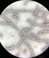
The negative staining procedure involves mixing bacteria with Nigrosin on a slide, spreading the mixture to form a thin smear, and air drying. The slide is then examined under oil immersion microscopy.
Label a clean slide with organism initials and your own.
Place a small drop of Nigrosin near the label.
Flame the rim of the culture tube and the loop.
Add a small amount of the organism to the Nigrosin drop and mix.
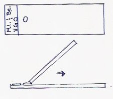
Use a second slide at a 30-degree angle to spread the mixture, forming a thin smear.
Allow the slide to air dry; do not heat fix or blot.
Examine under oil immersion and measure cell dimensions using an ocular micrometer.
Observing Microorganisms: Micrococcus luteus and Bacillus cereus
Micrococcus luteus
Micrococcus luteus is a Gram-negative coccus that forms tetrads and produces a yellow pigment. Its morphology and arrangement are best observed using negative staining.
Gram reaction: Gram-negative
Shape: Coccus (spherical)
Arrangement: Tetrads (groups of four)
Pigmentation: Yellow
Bacillus cereus
Bacillus cereus is a Gram-positive rod-shaped bacterium that often forms chains (strepto arrangement). Negative staining allows for accurate measurement of its length and width.
Gram reaction: Gram-positive
Shape: Bacillus (rod)
Arrangement: Chains (strepto)
Slide Preparation and Morphological Comparison
Slides can be prepared with individual species or a mixture to observe differences in morphology and arrangement. The following table summarizes the characteristics:
Slide | Organism | Arrangement | Shape |
|---|---|---|---|
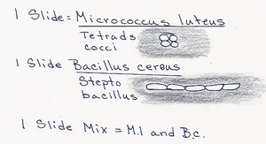
1 | Micrococcus luteus | Tetrads | Cocci |
2 | Bacillus cereus | Chains (strepto) | Bacilli |
3 | Mix (M.l. and B.c.) | Both | Cocci & Bacilli |
Measurement of Bacterial Dimensions
Using an Ocular Micrometer
To accurately determine the size of bacteria, measure the diameter of Micrococcus luteus and the length and width of Bacillus cereus using an ocular micrometer. Average measurements from approximately five cells and round off the result.
Diameter: Measure for cocci (e.g., M. luteus).
Length and width: Measure for bacilli (e.g., B. cereus).
Average: Calculate the mean value from multiple cells.
Summary Table: Negative Staining Characteristics
Stain Type | Charge | Cell Appearance | Background | Heat Fixation |
|---|---|---|---|---|
Negative (Nigrosin) | Negative | Transparent | Dark | No |
Positive (e.g., Crystal Violet) | Positive | Colored | Light | Yes |
Example: Negative staining is particularly useful for observing delicate structures such as capsules, which may be destroyed or distorted by heat fixation.
Additional info: Negative staining is a foundational technique in microbiology for studying cell morphology and arrangement, and is often used in conjunction with other staining methods for comprehensive analysis.
