 Back
BackProkaryotes: Domains Bacteria and Archaea – Structure, Classification, and Key Pathogens
Study Guide - Smart Notes
 Tailored notes based on your materials, expanded with key definitions, examples, and context.
Prokaryotes: Domains Bacteria and Archaea
Introduction to Prokaryotes
Prokaryotes are unicellular organisms lacking a membrane-bound nucleus and organelles. They are classified into two domains: Bacteria and Archaea. This module focuses on the diversity, structure, and clinical significance of bacteria, including their classification and representative pathogens.
Bacterial Morphology and Structure
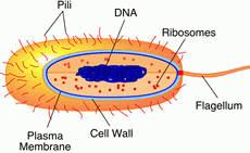
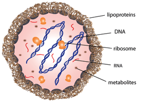
Basic Structure of Bacterial Cells
Bacteria exhibit a variety of shapes and structural features that are essential for their identification and function.
Cell Wall: Provides structural support and shape; composition varies between Gram-positive and Gram-negative bacteria.
Plasma Membrane: Regulates transport of substances into and out of the cell.
DNA: Typically a single, circular chromosome located in the nucleoid region.
Ribosomes: Sites of protein synthesis.
Pili: Hair-like structures involved in attachment and conjugation.
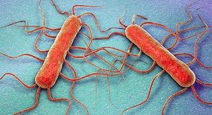
Flagellum: Provides motility.
Bacterial Morphologies
Bacteria can be classified by their shape and arrangement, which are important for identification in clinical microbiology.
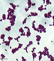
Cocci: Spherical bacteria (e.g., Staphylococcus, Streptococcus).
Bacilli: Rod-shaped bacteria (e.g., Escherichia coli, Bacillus).
Spirilla/Spirochetes: Spiral-shaped bacteria (e.g., Treponema).
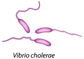
Vibrios: Comma-shaped bacteria (e.g., Vibrio cholerae).
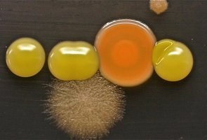
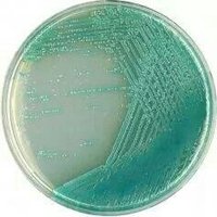
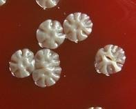
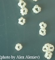
Bacterial Colonies on Agar Plates
Bacterial colonies display distinct morphologies on agar plates, which can aid in identification. Colony color, shape, and texture are diagnostic features.
Colony Morphology: Includes size, shape, color, and surface appearance.
Mixed Cultures: Plates may contain both bacterial and fungal colonies.
Classification of Bacteria
Proteobacteria
Proteobacteria is the largest phylum of Gram-negative bacteria, encompassing a wide variety of pathogens and environmental organisms. They are classified into several classes based on genetic and metabolic characteristics.
Gram-negative: Characterized by a thin peptidoglycan layer and an outer membrane.
Chemoheterotrophic: Obtain energy by consuming organic molecules.
Major Classes: Alphaproteobacteria, Betaproteobacteria, Gammaproteobacteria, Epsilonproteobacteria.
Alphaproteobacteria
Rickettsia: Obligate intracellular parasites; cause spotted fevers (e.g., R. rickettsii – Rocky Mountain spotted fever).
Rhizobium/Bradyrhizobium: Nitrogen-fixing bacteria in legume roots.
Agrobacterium: Plant pathogen; used in biotechnology for genetic engineering.
Bartonella: Causes cat-scratch disease.
Brucella: Causes brucellosis; survives phagocytosis.
Betaproteobacteria
Burkholderia: Degrades organic molecules; some species are opportunistic pathogens.
Bordetella pertussis: Causes whooping cough.
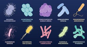
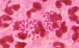
Neisseria: Includes N. gonorrhoeae (gonorrhea) and N. meningitidis (meningococcal meningitis).
Gammaproteobacteria
Pseudomonas: Opportunistic pathogens, common in soil, resistant to antibiotics (e.g., P. aeruginosa).
Legionella: Causes Legionnaire’s disease; found in water systems.
Vibrio: Curved rods; V. cholerae causes cholera.
Enterobacteriales (Enterics): Inhabit intestinal tract; ferment carbohydrates; include Escherichia, Salmonella, Shigella, Klebsiella, Serratia, Proteus, Yersinia, Enterobacter.

Pasteurellales: Includes Pasteurella (animal pathogen) and Haemophilus (requires X and V factors for growth; causes meningitis, pneumonia).
Epsilonproteobacteria
Campylobacter: Causes foodborne intestinal disease.
Helicobacter pylori: Causes peptic ulcers and stomach cancer.
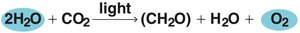
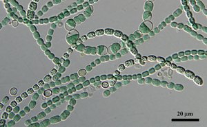
Cyanobacteria (Photosynthetic Bacteria)
Cyanobacteria, also known as blue-green algae, perform oxygenic photosynthesis and are important for nitrogen fixation in aquatic environments.
Photosynthesis Equation:
Heterocysts: Specialized cells for nitrogen fixation.
Gas Vesicles: Provide buoyancy.
Unicellular or Filamentous: Example: Anabaena species.
Chlamydiae
Chlamydiae are obligate intracellular bacteria lacking peptidoglycan in their cell walls. They form infectious elementary bodies and cause a variety of diseases.
Chlamydia trachomatis: Causes trachoma and urethritis.
Chlamydophila psittaci: Causes respiratory psittacosis.
Chlamydophila pneumoniae: Causes mild pneumonia in young adults.
Nonproteobacteria Gram-Negative Bacteria
Bacteroidetes: Anaerobic; found in mouth and large intestine.
Fusobacteria: Anaerobic; found in mouth; cause dental abscesses.
Spirochaetes
Spirochaetes are coiled bacteria that move via axial filaments. They include several important human pathogens.
Treponema pallidum: Causes syphilis (STD).
Borrelia: Causes relapsing fever and Lyme disease.
Leptospira: Excreted in animal urine; causes leptospirosis.

Deinococci
Deinococcus radiodurans: Highly resistant to radiation.
Thermus aquaticus: Source of Taq polymerase for PCR.
Gram-Positive Bacteria
Firmicutes (Low G + C Gram-Positive Bacteria)
Clostridium: Endospore-producing obligate anaerobes; includes C. tetani, C. botulinum, C. perfringens, C. difficile.
Bacillus: Endospore-producing rods; includes B. anthracis (anthrax), B. cereus (food poisoning).
Staphylococcus: Grape-like clusters; S. aureus causes wound infections, is often antibiotic resistant, and produces enterotoxins.
Lactobacillus: Aerotolerant anaerobes; used in food production.
Streptococcus: Spherical in chains; includes beta-hemolytic (S. pyogenes) and non-beta-hemolytic (S. mutans, S. pneumoniae).
Enterococcus: Found in intestinal tract; can infect surgical wounds and urinary tract.
Listeria monocytogenes: Food contaminant; can cause listeriosis.
Mycoplasma: Lack cell wall; pleomorphic; M. pneumoniae causes mild pneumonia.
Actinobacteria (High G + C Gram-Positive Bacteria)
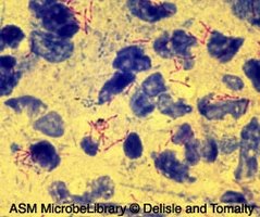
Mycobacterium: Waxy, acid-fast cell wall; includes M. tuberculosis (tuberculosis) and M. leprae (leprosy).
Corynebacterium diphtheriae: Causes diphtheria.
Propionibacterium acnes: Causes acne.
Gardnerella vaginalis: Causes vaginitis.
Streptomyces: Soil bacteria; produce most antibiotics.
Actinomyces: Form filaments in mouth and throat; destroy tissues.
Nocardia: Acid-fast; causes pulmonary infections.
Uncultured and Environmental Bacteria
Metagenomics and Bacterial Diversity
Modern molecular techniques such as PCR and metagenomics have revealed a vast diversity of bacteria in the environment, many of which have not been cultured. DNA barcoding is used to identify these organisms and understand their ecological roles.
Metagenomics: Analysis of genetic material recovered directly from environmental samples.
Barcoding: Uses short genetic sequences for rapid identification.
Additional info: These advances are crucial for studying complex microbial communities and their impact on health and the environment.
