Back
BackProkaryotic Cell Structure and Function: Study Notes for Microbiology
Study Guide - Smart Notes
 Tailored notes based on your materials, expanded with key definitions, examples, and context.
Prokaryotic Cell Basics
Domains of Prokaryotes
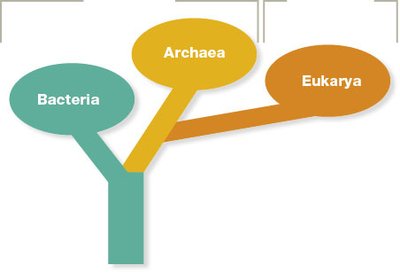
Prokaryotic cells are classified into two major domains: Bacteria and Archaea. Both domains consist of organisms lacking a membrane-bound nucleus and organelles, but they differ in several molecular and structural features.
Bacteria: Characterized by peptidoglycan in their cell walls and unique ribosomal RNA sequences.
Archaea: Possess pseudopeptidoglycan or other polymers in their cell walls and have distinct membrane lipids.
Both domains share a common ancestor with Eukarya, the domain containing all eukaryotic organisms.
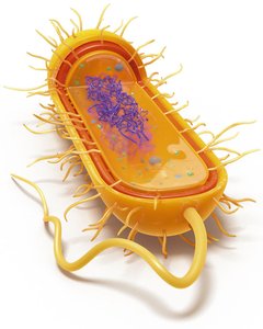
Basic Structure of Prokaryotic Cells
Prokaryotic cells are generally small (0.2–2.0 μm in diameter) and lack a true nucleus. Their small size allows for efficient nutrient uptake and waste removal, supporting rapid growth and division.
Cytoplasm: Gel-like substance containing water, enzymes, nutrients, wastes, and gases.
Nucleoid: Region containing the cell’s circular DNA chromosome.
Ribosomes: Sites of protein synthesis.
Plasma membrane: Phospholipid bilayer controlling entry and exit of substances.
Cell wall: Provides structural support and shape.
Capsule (in some): Protective, sticky outer layer.
Fimbriae and flagella (in some): Structures for attachment and motility.
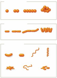
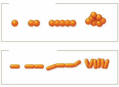
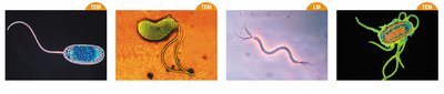
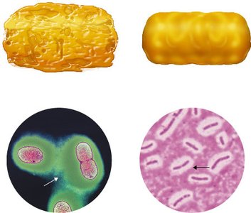
Shapes and Arrangements of Prokaryotes
Prokaryotes exhibit a variety of shapes and arrangements, which are important for identification and classification.
Bacilli: Rod-shaped
Cocci: Spherical
Other shapes: Vibrio (comma-shaped), Spirochete (spiral), Coccobacillus (short rod), Stella (star-shaped), Filamentous, Pleomorphic (variable shape)
Arrangements result from cell division patterns:
Diplo-: Pairs
Strepto-: Chains
Staphylo-: Clusters (cocci only)
Palisades: Side-by-side arrangement (bacilli)
Pleomorphism
Pleomorphic organisms can alter their shape or size in response to environmental conditions. This adaptability may enhance their ability to evade the immune system or survive in diverse environments.
Binary Fission
Prokaryotic cells reproduce asexually by binary fission, a process involving:
Replication of the DNA chromosome
Elongation of the cell
Formation of a septum (partition) at the cell midpoint
Division into two genetically identical daughter cells
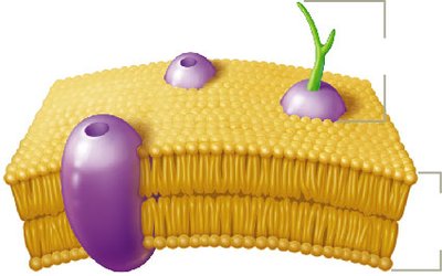
Extracellular Structures of Prokaryotes
Plasma Membrane Structure and Function
The plasma membrane is a thin, flexible barrier composed of a phospholipid bilayer with embedded proteins. It acts as a selective barrier, regulating the movement of substances in and out of the cell. Membrane fluidity is influenced by temperature and fatty acid composition.
Bacteria: Linear fatty acids in phospholipids
Archaea: Branched, sometimes monolayer-forming fatty acids for stability in extreme environments
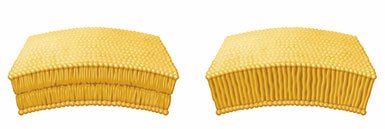
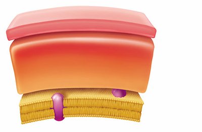
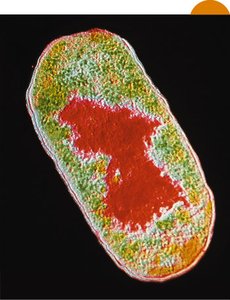
Cell Wall Composition
The cell wall provides rigidity and protection. Its composition differs between domains:
Bacteria: Peptidoglycan (a polymer of sugars and amino acids)
Archaea: Pseudopeptidoglycan or other polymers
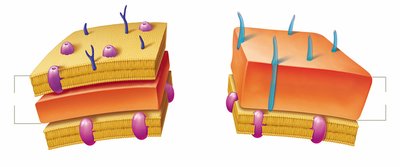
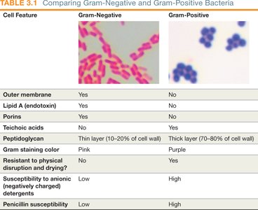
Gram-Positive vs. Gram-Negative Bacteria
Bacteria are classified based on cell wall structure, which affects their Gram stain reaction and clinical properties.
Feature | Gram-Negative | Gram-Positive |
|---|---|---|
Outer membrane | Yes | No |
Lipid A (endotoxin) | Yes | No |
Porins | Yes | No |
Teichoic acids | No | Yes |
Peptidoglycan | Thin (10–20%) | Thick (70–80%) |
Gram staining color | Pink | Purple |
Physical resistance | No | Yes |
Detergent susceptibility | Low | High |
Penicillin susceptibility | Low | High |
Acid-Fast Bacteria
Some bacteria, such as Mycobacterium and Nocardia, have waxy mycolic acid in their cell walls, making them resistant to Gram staining. Acid-fast staining is used to identify these clinically important pathogens.
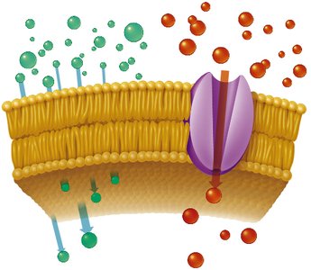
Passive and Active Transport Mechanisms
Cells transport substances across membranes using passive and active mechanisms:
Passive transport: Does not require energy. Includes simple diffusion, facilitated diffusion, and osmosis.
Active transport: Requires energy (usually ATP) to move substances against their concentration gradients.
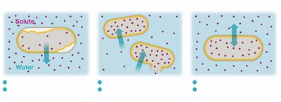
Osmosis and Tonicity
Osmosis is the diffusion of water across a selectively permeable membrane. The effect of different environments on bacterial cells:
Hypertonic: Water leaves the cell, causing plasmolysis.
Hypotonic: Water enters the cell, possibly causing lysis if the cell wall is damaged.
Isotonic: No net water movement; cell remains stable.
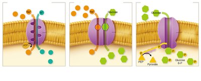
Active Transport Types
Primary active transport: Direct use of ATP (e.g., sodium-potassium pump).
Secondary active transport: Uses the energy from an ion gradient (e.g., symporters).
Phosphotransferase system: Chemically modifies the transported substance (e.g., glucose phosphorylation).
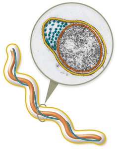
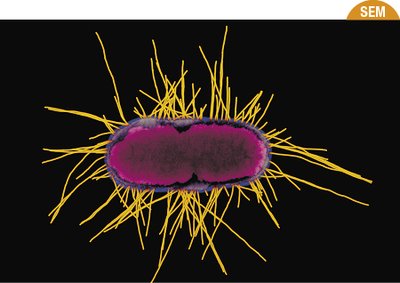
Flagella
Flagella are long, filamentous structures used for motility. They are composed of the protein flagellin and are anchored in the cell wall and membrane by a basal body.
Enable movement via a run-and-tumble mechanism.
Arrangement varies: monotrichous (single), lophotrichous (tuft), amphitrichous (both poles), peritrichous (all over).

Periplasmic Flagella (Axial Filaments)
Found in spirochetes, these flagella are located between the plasma membrane and cell wall, allowing corkscrew movement.
Fimbriae and Pili
Fimbriae: Short, bristle-like structures for adhesion and biofilm formation; common in Gram-negative bacteria.
Pili: Longer, less numerous; involved in adhesion, movement, and gene transfer (conjugation).
Glycocalyx
The glycocalyx is a sticky, carbohydrate-rich layer outside the cell wall. It can be a loosely organized slime layer or a well-organized capsule, aiding in protection and adherence.
Intracellular Structures of Prokaryotes
Nucleoid
The nucleoid is the region where the prokaryotic chromosome (usually a single, circular DNA molecule) is located. It is not surrounded by a membrane.
Ribosomes
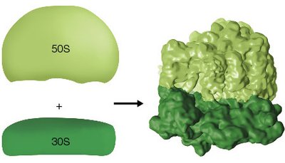
Prokaryotic ribosomes (70S) are composed of a large (50S) and a small (30S) subunit. They are the sites of protein synthesis and are structurally distinct from eukaryotic ribosomes, supporting the endosymbiotic theory.
Cytoskeleton
The prokaryotic cytoskeleton consists of long protein filaments that provide structural support and help maintain cell shape.
Inclusion Bodies
Inclusion bodies are storage sites for nutrients and other substances. Examples include:
Carboxysomes: Contain enzymes for carbon fixation.
Magnetosomes: Contain magnetic iron for orientation in magnetic fields.
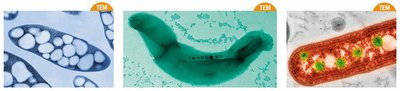
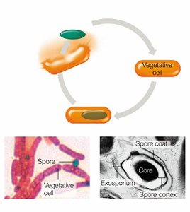
Endospores
Endospores are metabolically inactive, highly resistant structures formed by certain bacteria (e.g., Bacillus, Clostridium) in response to stress. They can survive extreme conditions and are a concern in healthcare due to their persistence and resistance to disinfection.
Sporulation: The process of endospore formation, involving DNA replication, packaging, and formation of protective layers.
Endospores germinate into vegetative cells when conditions improve.