 Back
BackProkaryotic Cell Structure, Morphology, and Staining in Microbiology
Study Guide - Smart Notes
 Tailored notes based on your materials, expanded with key definitions, examples, and context.
Prokaryotic Cell Morphology and Structure
Introduction to Prokaryotes
Prokaryotes are unicellular organisms that lack a membrane-bound nucleus and organelles. They include bacteria and archaea, which are fundamental to the study of microbiology due to their diversity, ecological roles, and medical importance.
Cell Morphology: Prokaryotes exhibit various shapes, including cocci (spherical), bacilli (rod-shaped), spirilla (spiral), vibrios (comma-shaped), and pleomorphic forms.
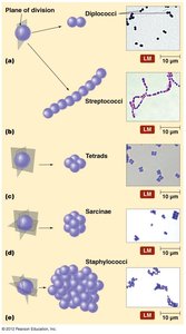
Cell Arrangements: Cells may occur singly or in characteristic arrangements such as chains (strepto-), clusters (staphylo-), pairs (diplo-), tetrads, or sarcinae, depending on the plane of division and cellular adhesion.
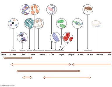
Size Range: Prokaryotic cells are typically 0.2–2.0 µm in diameter, much smaller than most eukaryotic cells.
Comparison: Prokaryotes lack membrane-bound organelles, while eukaryotes possess compartmentalized structures such as nuclei and mitochondria.
Common Prokaryotic Shapes and Arrangements
Cocci: Spherical bacteria that may form diplococci, streptococci, tetrads, sarcinae, or staphylococci arrangements.
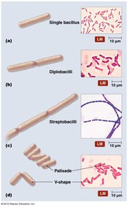
Bacilli: Rod-shaped bacteria that may appear as single bacilli, diplobacilli, streptobacilli, or palisades.
Other Shapes: Vibrio (curved rods), spirillum (rigid spiral), spirochete (flexible spiral), and pleomorphic (variable shape).
Examples of Pathogenic Bacteria
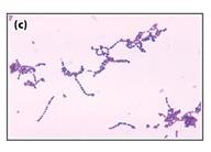
Streptococcus pyogenes: Causes strep throat; appears as chains of cocci.
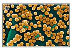
Staphylococcus aureus: Causes pneumonia and other infections; appears as clusters of cocci.
Staining Techniques in Microbiology
Purpose of Staining
Staining enhances the contrast of microbial cells under the microscope, allowing for visualization of cell morphology, arrangement, and structural features. Different stains are used for specific diagnostic and research purposes.
Types of Stains
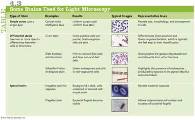
Type of Stain | Examples | Results | Representative Uses |
|---|---|---|---|
Simple stains | Crystal violet, methylene blue | Uniform purple or blue stain | Reveals size, morphology, and arrangement of cells |
Gram stain | Crystal violet, iodine, alcohol, safranin | Gram-positive: purple/blue Gram-negative: pink/red | Differentiates Gram-positive and Gram-negative bacteria |
Acid-fast stain | Ziehl-Neelsen | Acid-fast cells: pink/red Non-acid-fast: blue | Detects Mycobacterium and Nocardia |
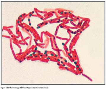
Endospore stain | Schaeffer-Fulton | Endospores: green Cells: pink/red | Highlights endospores in Bacillus and Clostridium |
Negative stain | India ink, nigrosin | Background is dark, cells are unstained | Reveals capsules |
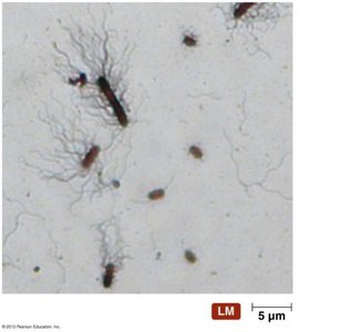
Flagella stain | Carbolfuchsin with mordant | Bacterial flagella become visible | Determines number and location of flagella |
Gram Stain Procedure and Interpretation
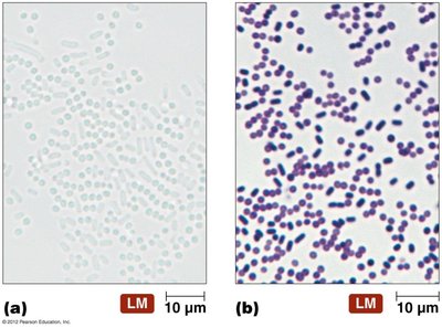
Principle: Differentiates bacteria based on cell wall structure.
Steps: Application of crystal violet, iodine (mordant), alcohol (decolorizer), and safranin (counterstain).
Results: Gram-positive bacteria retain crystal violet (purple/blue); Gram-negative bacteria lose crystal violet and take up safranin (pink/red).
Clinical Importance: First step in bacterial identification in clinical labs.
Special Stains
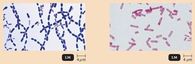
Acid-fast stain: Detects mycolic acid in cell walls of Mycobacterium species (e.g., M. tuberculosis).
Endospore stain: Identifies endospores in genera such as Bacillus and Clostridium.
Capsule stain: Visualizes the protective capsule surrounding some bacteria.
Flagella stain: Reveals the presence and arrangement of flagella.
Prokaryotic Cell Structure and Function
Overview of Bacterial Cell Structure
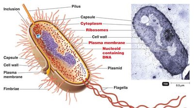
Bacterial cells are surrounded by a cell envelope, which includes the plasma membrane, cell wall, and sometimes a capsule. The cytoplasm contains the nucleoid, ribosomes, and various inclusions.
The Plasma Membrane
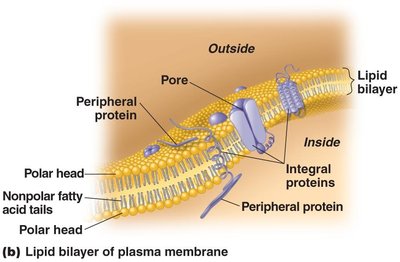
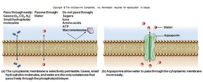
Fluid Mosaic Model: The plasma membrane is a selectively permeable phospholipid bilayer with embedded proteins.
Functions: Acts as a barrier, mediates transport, and contains proteins for energy generation and signal transduction.
Selective Permeability and Transport Mechanisms
Simple Diffusion: Movement of small, nonpolar molecules down their concentration gradient.
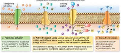
Facilitated Diffusion: Transport of substances via membrane proteins down their concentration gradient.
Active Transport: Movement of substances against their concentration gradient using energy (ATP or proton motive force).
Group Translocation: Substance is chemically modified during transport (e.g., phosphotransferase system).
Osmosis and the Cell Wall
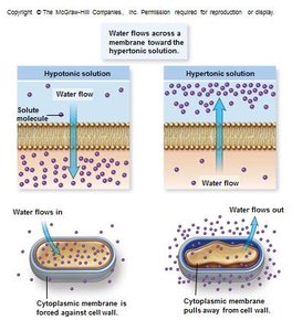
Osmosis: Movement of water across a selectively permeable membrane from low to high solute concentration.
Cell Wall Function: Protects against osmotic lysis by providing structural support.
Cell Wall Structure: Gram-Positive vs. Gram-Negative Bacteria
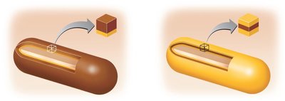
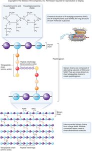
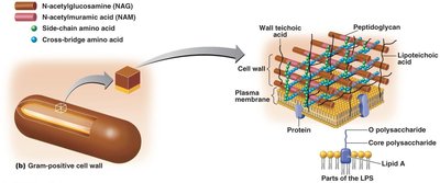
Peptidoglycan: Main component of bacterial cell walls, consisting of glycan chains cross-linked by peptides.
Gram-Positive: Thick peptidoglycan layer, teichoic acids, no outer membrane.
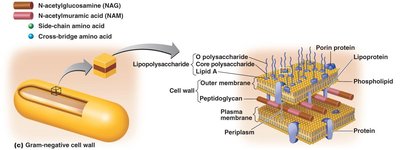
Gram-Negative: Thin peptidoglycan layer, outer membrane with lipopolysaccharide (LPS), periplasmic space.
LPS: Contains Lipid A (endotoxin) and O polysaccharide (diagnostic marker).
Specialized Cell Wall Types
Acid-Fast Cell Walls: Contain mycolic acid; characteristic of Mycobacterium species.
Atypical Cell Walls: Some bacteria lack peptidoglycan or have unique cell wall components.
Glycocalyx: Capsule and Slime Layer
Capsule: Organized, firmly attached layer; protects against desiccation and phagocytosis, aids in adherence and biofilm formation.
Slime Layer: Unorganized, loosely attached layer; also aids in adherence.
Composition: Polysaccharides, polypeptides, or both.
Motility Structures: Flagella, Fimbriae, and Pili
Flagella: Long, whip-like structures for motility; composed of filament, hook, and basal body.
Arrangements: Monotrichous, lophotrichous, amphitrichous, peritrichous, and axial filaments (spirochetes).
Chemotaxis: Movement toward or away from chemical stimuli.
Fimbriae: Short, hair-like projections for attachment and biofilm formation.
Pili: Longer than fimbriae; involved in motility and genetic exchange (conjugation).
Internal Structures: Nucleoid, Plasmids, Ribosomes, and Inclusions
Nucleoid: Region containing the single, circular, double-stranded DNA chromosome (not membrane-bound).
Plasmids: Small, circular DNA molecules; often carry antibiotic resistance genes.
Ribosomes: Sites of protein synthesis; prokaryotic ribosomes are 70S (50S + 30S subunits).
Inclusions: Storage granules for nutrients such as glycogen, poly-β-hydroxybutyrate (PHB), phosphate, sulfur, or nitrogen.
Endospores
Definition: Highly resistant, dormant structures formed by some bacteria (e.g., Bacillus, Clostridium) for survival under adverse conditions.
Resistance: Withstand heat, radiation, chemicals, desiccation, and boiling water.
Structure: Dehydrated core with DNA-binding proteins (SASPs), surrounded by cortex, spore coat, and exosporium; contains calcium dipicolinate for protection.
Sporulation: Process of endospore formation; germination returns the spore to a vegetative cell.
Comparison: Prokaryotic vs. Eukaryotic Cells
Prokaryotes: No membrane-bound nucleus or organelles; single circular chromosome; 70S ribosomes; cell wall usually contains peptidoglycan.
Eukaryotes: Membrane-bound nucleus and organelles; multiple linear chromosomes; 80S ribosomes; cell wall (if present) lacks peptidoglycan.
Additional info: This guide covers foundational concepts in prokaryotic cell structure, morphology, and staining, which are essential for understanding microbial taxonomy, physiology, and clinical identification.
