 Back
BackChapter Five
Study Guide - Smart Notes
 Tailored notes based on your materials, expanded with key definitions, examples, and context.
Viruses: Structure, Replication, and Diversity
What Is a Virus?
Viruses are obligate intracellular parasites that require a host cell to replicate. They possess their own genetic material, which can be single-stranded (ss) or double-stranded (ds) RNA or DNA. The virion is the extracellular, infectious form of the virus. Not all viruses cause disease; some may exist in a host without causing noticeable effects.
Genome: Can be DNA or RNA, single- or double-stranded.
Virion: The complete, infectious virus particle outside a host cell.
Obligate parasite: Cannot reproduce independently; must infect a host cell.
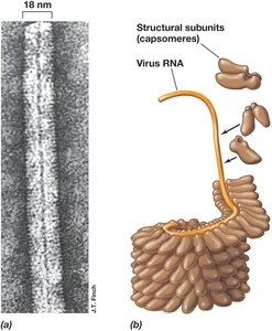
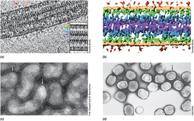
Structure of the Virion
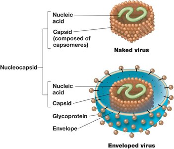
The virion consists of a nucleic acid genome surrounded by a protein coat called the capsid. Some viruses also possess an outer lipid envelope derived from the host cell membrane. The capsid is made up of protein subunits called capsomeres. Viruses exhibit a variety of shapes, including helical, icosahedral, and complex forms.
Naked virus: Contains only nucleic acid and capsid.
Enveloped virus: Has an additional lipid membrane surrounding the capsid.
Capsomeres: Protein subunits that make up the capsid.
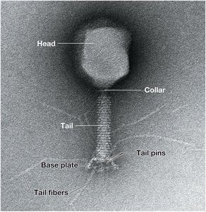
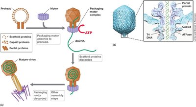
Complex Virus Structures
Some viruses, such as bacteriophages, have complex structures with specialized components for host recognition and genome delivery. These may include a head (capsid), tail, base plate, and tail fibers.
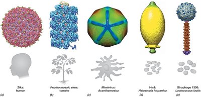
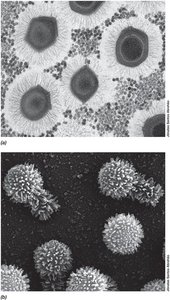
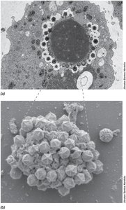
Virion Morphology and Diversity
Viruses display a wide range of morphologies, from simple helical and icosahedral forms to more elaborate structures. Electron microscopy reveals details of viral architecture, including surface projections and internal organization.
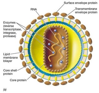
Enveloped Viruses
Enveloped viruses, such as retroviruses, possess a lipid bilayer derived from the host cell membrane. This envelope contains viral glycoproteins essential for host cell recognition and entry. The core contains the viral genome and associated proteins, including enzymes necessary for replication.
Envelope: Lipid bilayer with embedded viral proteins.
Core: Contains viral genome and enzymes (e.g., reverse transcriptase in retroviruses).
Culturing, Detecting, and Counting Viruses
Methods for Virus Quantification
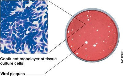
Viruses are typically cultured in living cells, such as bacterial lawns or tissue cultures. The presence of viruses can be detected by the formation of plaques, which are clear zones resulting from cell lysis. Plaque assays are a standard method for quantifying infectious virus particles.
Plaque: Clear area on a cell layer caused by virus-induced cell lysis.
Plaque-forming unit (PFU): A measure of infectious virus quantity.
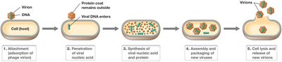
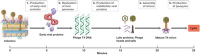
Replication Cycle of Lytic Bacterial Viruses
Lytic Cycle Steps
The lytic replication cycle of bacteriophages involves several distinct steps:
Attachment (adsorption): The phage binds to specific receptors on the host cell surface.
Penetration: The viral genome enters the host cell, while the protein coat remains outside.
Synthesis: Host machinery is redirected to synthesize viral nucleic acids and proteins.
Assembly: New virions are assembled from synthesized components.
Release: Host cell lysis releases new virions.
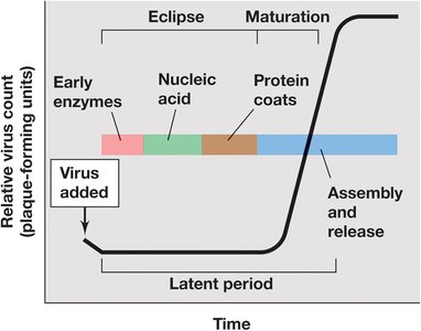
One-Step Growth Curve of Viral Replication
The one-step growth curve illustrates the replication dynamics of viruses in a host population. It includes the eclipse period (no infectious particles detected), followed by a rise in virus count as new virions are assembled and released.
Eclipse period: Time during which viral components are being synthesized but no complete virions are present.
Latent period: Time from infection to the release of new virions.
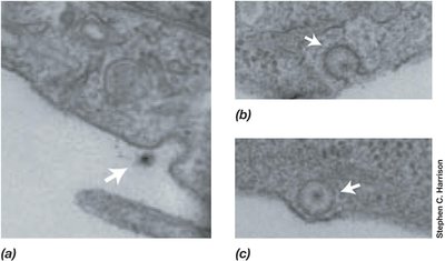
Host Recognition and Attachment
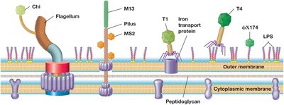
Coliphage Receptors on E. coli
Bacteriophages recognize and bind to specific receptors on the bacterial surface, such as flagella, pili, or membrane proteins. This specificity determines the host range of the phage.
Attachment and Infection of E. coli by Phage T4
Phage T4 attaches to the surface of E. coli using tail fibers, then injects its DNA into the host cytoplasm. The tail contracts, and the viral genome is delivered through the cell envelope.
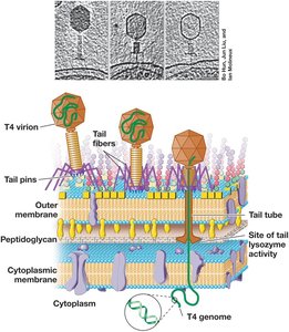
Bacteriophage T4: A Model Lytic Virus
Bacteriophage T4 is a well-studied model for understanding lytic viral replication. Its infection process includes early and late gene expression, DNA replication, assembly of head and tail structures, and host cell lysis.
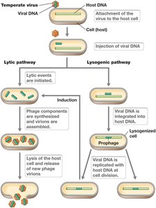
Temperate Bacteriophages and Lysogeny
Lytic vs. Lysogenic Pathways
Temperate phages can undergo two types of replication cycles:
Lytic pathway: Phage replicates and lyses the host cell.
Lysogenic pathway: Phage DNA integrates into the host genome as a prophage and replicates with the host without causing immediate lysis. Induction can trigger entry into the lytic cycle.
Viruses of Eukaryotes
Overview of Eukaryotic Viruses
Viruses infecting eukaryotes include those that target animals, plants, and protists. They may cause acute, persistent, or latent infections, and some can transform host cells, leading to tumor formation.
Acute infection: Rapid onset and resolution.
Persistent infection: Virus remains in the host for extended periods.
Latent infection: Virus is present but not actively replicating.
Transformation: Virus induces uncontrolled cell division (tumor formation).
Viral Infection of Plants
Plant viruses can have DNA or RNA genomes and infect a wide range of species. They are often transmitted by insect vectors or mechanical damage. Examples include Cauliflower mosaic virus (dsDNA), Tomato spotted wilt virus (ssRNA), and Rice black streaked dwarf virus (dsRNA).
Disease or Host | Virus | Genome | Size (nt) |
|---|---|---|---|
Cauliflower, turnip, cabbage | Cauliflower mosaic virus | dsDNA | 8,000 |
Cassava | African cassava virus | ssDNA | 5,500 |
Strawberry | Strawberry crinkle virus | ssRNA | 14,500 |
>900 plant species | Tomato spotted wilt virus | ssRNA | 16,600 |
Tobacco, tomato, pepper | Tobacco mosaic virus | ssRNA | 6,500 |
>1200 plant species | Cucumber mosaic virus | ssRNA | 8,600 |
Rice, maize, wheat, barley | Rice black streaked dwarf virus | dsRNA | 29,100 |
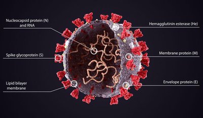
Generalized Structure of Coronavirus
Coronaviruses are enveloped, positive-sense ssRNA viruses with a characteristic crown-like appearance due to spike glycoproteins. The genome is enclosed by a nucleocapsid protein, and the envelope contains several structural proteins, including spike (S), membrane (M), envelope (E), and hemagglutinin esterase (He).
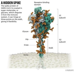
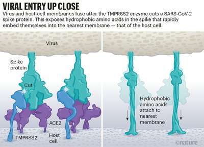
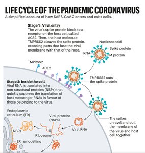
Attachment of SARS-CoV-2
The spike protein of SARS-CoV-2 binds to the ACE2 receptor on host cells. The spike protein is heavily glycosylated, which helps it evade the immune system. After binding, host proteases (such as TMPRSS2) cleave the spike protein, facilitating membrane fusion and viral entry.
Syncytia Formation in SARS-CoV-2 Infection
Cells expressing the SARS-CoV-2 spike protein can fuse to form multinucleated structures called syncytia. This process is mediated by the spike protein and may contribute to viral spread and pathogenesis within tissues.
Additional info: The study of viruses is essential for understanding infectious diseases, biotechnology applications, and the molecular mechanisms of host-pathogen interactions. Viruses are also important tools in genetic engineering and gene therapy research.
