Textbook Question
Which of the following is not a modification of a compound light microscope?
a. Brightfield microscopy
b. Darkfield microscopy
c. Electron microscopy
d. Phase-contrast microscopy
e. Fluorescence microscopy
1449
views
 Verified step by step guidance
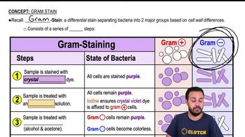
 04:57
04:57 03:44 04:57
Which of the following is not a modification of a compound light microscope?
a. Brightfield microscopy
b. Darkfield microscopy
c. Electron microscopy
d. Phase-contrast microscopy
e. Fluorescence microscopy
Assume that you are viewing a Gram-stained field of red cocci and blue rods through the microscope. You can safely conclude that you have
a. Made a mistake in staining
b. Two different species
c. Old bacterial cells
d. Young bacterial cells
e. None of the above
In 1996, scientists described a new tapeworm parasite that had killed at least one person. The initial examination of the patient’s abdominal mass was most likely made using:
a. Brightfield microscopy
b. Darkfield microscopy
c. Electron microscopy
d. Phase-contrast microscopy
e. Fluorescence microscopy
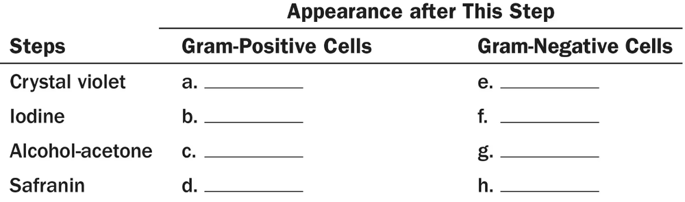
What is the purpose of a decolorizer in the Gram stain? In the acid-fast stain?